 Movie
Movie Controller


+ Open data

- Basic information
| Entry | Database: PDB / ID: 8dj6 | ||||||
|---|---|---|---|---|---|---|---|
| Title | Sliding-clamp-ImuB peptide | ||||||
 Components Components |
| ||||||
 Keywords Keywords | DNA BINDING PROTEIN / Sliding clamp in complex with ImuB peptide | ||||||
| Function / homology |  Function and homology information Function and homology informationDNA polymerase III complex / DNA strand elongation involved in DNA replication / 3'-5' exonuclease activity / DNA-directed DNA polymerase activity / DNA binding / identical protein binding / cytoplasm Similarity search - Function | ||||||
| Biological species |  Mycolicibacterium thermoresistibile ATCC 19527 (bacteria) Mycolicibacterium thermoresistibile ATCC 19527 (bacteria) | ||||||
| Method |  X-RAY DIFFRACTION / SYNCHROTRON / MOLECULAR REPLACEMENT / Resolution: 2.5 Å X-RAY DIFFRACTION / SYNCHROTRON / MOLECULAR REPLACEMENT / Resolution: 2.5 Å | ||||||
 Authors Authors | Kapur, M.K. / Gray, O.J. / Honzatko, R.H. / Nelson, S.N. | ||||||
| Funding support |  United States, 1items United States, 1items
| ||||||
 Citation Citation | Journal: To Be Published Title: Interaction of sliding clamp with mycobacterial polymerases Authors: Kapur, M.K. / Gray, O.J. / Honzatko, R.H. / Nelson, S.N. | ||||||
| History |
|
- Structure visualization
| Structure viewer | Molecule: MolmilJmol/JSmol |
|---|
- Downloads & links
-Download
| PDBx/mmCIF format | 8dj6.cif.gz | 604.7 KB | Display | PDBx/mmCIF format |
|---|---|---|---|---|
| PDB format | pdb8dj6.ent.gz | 470 KB | Display | PDB format |
| PDBx/mmJSON format | 8dj6.json.gz | Tree view | PDBx/mmJSON format | |
| Others |  Other downloads Other downloads |
-Validation report
| Arichive directory | https://data.pdbj.org/pub/pdb/validation_reports/dj/8dj6ftp://data.pdbj.org/pub/pdb/validation_reports/dj/8dj6 | HTTPS FTP |
|---|
-Related structure data
| Related structure data |  8dk9C 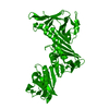 8dkdC  4tr7S S: Starting model for refinement C: citing same article ( |
|---|---|
| Similar structure data |
-Links
 PDBj
PDBj
- Assembly
| Deposited unit | 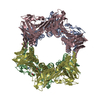
| ||||||||||||
|---|---|---|---|---|---|---|---|---|---|---|---|---|---|
| 1 | 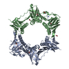
| ||||||||||||
| 2 | 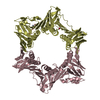
| ||||||||||||
| Unit cell |
|
-Components
| #1: Protein | Mass: 41677.191 Da / Num. of mol.: 4 Source method: isolated from a genetically manipulated source Source: (gene. exp.) Mycolicibacterium thermoresistibile ATCC 19527 (bacteria)Strain: ATCC 19527 / DSM 44167 / CIP 105390 / JCM 6362 / NCTC 10409 / 316 Gene: KEK_10778 / Production host: #2: Protein/peptide | Mass: 736.882 Da / Num. of mol.: 4 / Source method: obtained synthetically Source: (synth.) Mycolicibacterium thermoresistibile ATCC 19527 (bacteria)#3: Chemical | ChemComp-FMT /   Mass: 46.025 Da / Num. of mol.: 7 / Source method: obtained synthetically / Formula: CH2O2 Mass: 46.025 Da / Num. of mol.: 7 / Source method: obtained synthetically / Formula: CH2O2#4: Chemical | ChemComp-ACT / |   Mass: 59.044 Da / Num. of mol.: 1 / Source method: obtained synthetically / Formula: C2H3O2 Mass: 59.044 Da / Num. of mol.: 1 / Source method: obtained synthetically / Formula: C2H3O2#5: Water | ChemComp-HOH / |  Mass: 18.015 Da / Num. of mol.: 371 / Source method: isolated from a natural source / Formula: H2O Mass: 18.015 Da / Num. of mol.: 371 / Source method: isolated from a natural source / Formula: H2OHas ligand of interest | Y | Has protein modification | Y | |
|---|
-Experimental details
-Experiment
| Experiment | Method: X-RAY DIFFRACTION / Number of used crystals: 1 |
|---|
- Sample preparation
| Crystal | Density Matthews: 4.31 Å3/Da / Density % sol: 71.45 % |
|---|---|
| Crystal grow | Temperature: 291.5 K / Method: vapor diffusion, sitting drop / Details: 0.1M Bis -Tris pH6.3, 60% Tascimate pH7 |
-Data collection
| Diffraction | Mean temperature: 100 K / Serial crystal experiment: N |
|---|---|
| Diffraction source | Source: SYNCHROTRON / Site: APS / Beamline: 23-ID-B / Wavelength: 1 Å |
| Detector | Type: DECTRIS PILATUS3 6M / Detector: PIXEL / Date: Jul 22, 2019 |
| Radiation | Protocol: SINGLE WAVELENGTH / Monochromatic (M) / Laue (L): M / Scattering type: x-ray |
| Radiation wavelength | Wavelength: 1 Å / Relative weight: 1 |
| Reflection | Resolution: 2.5→46.75 Å / Num. obs: 94524 / % possible obs: 96.4 % / Redundancy: 3 % / Biso Wilson estimate: 54.86 Å2 / CC1/2: 0.963 / Rmerge(I) obs: 0.123 / Rpim(I) all: 0.082 / Net I/σ(I): 10.3 |
| Reflection shell | Resolution: 2.5→2.54 Å / Rmerge(I) obs: 0.988 / Mean I/σ(I) obs: 0.88 / Num. unique obs: 4590 / CC1/2: 0.4 / Rpim(I) all: 0.671 |
- Processing
| Software |
| |||||||||||||||||||||||||||||||||||||||||||||||||||||||||||||||||||||||||||||||||||||||||||||||||||||||||
|---|---|---|---|---|---|---|---|---|---|---|---|---|---|---|---|---|---|---|---|---|---|---|---|---|---|---|---|---|---|---|---|---|---|---|---|---|---|---|---|---|---|---|---|---|---|---|---|---|---|---|---|---|---|---|---|---|---|---|---|---|---|---|---|---|---|---|---|---|---|---|---|---|---|---|---|---|---|---|---|---|---|---|---|---|---|---|---|---|---|---|---|---|---|---|---|---|---|---|---|---|---|---|---|---|---|---|
| Refinement | Method to determine structure: MOLECULAR REPLACEMENT Starting model: 4TR7 Resolution: 2.5→46.75 Å / SU ML: 0.3461 / Cross valid method: FREE R-VALUE / σ(F): 1.36 / Phase error: 26.0738 Stereochemistry target values: GeoStd + Monomer Library + CDL v1.2
| |||||||||||||||||||||||||||||||||||||||||||||||||||||||||||||||||||||||||||||||||||||||||||||||||||||||||
| Solvent computation | Shrinkage radii: 0.9 Å / VDW probe radii: 1.11 Å / Solvent model: FLAT BULK SOLVENT MODEL | |||||||||||||||||||||||||||||||||||||||||||||||||||||||||||||||||||||||||||||||||||||||||||||||||||||||||
| Displacement parameters | Biso mean: 64.01 Å2 | |||||||||||||||||||||||||||||||||||||||||||||||||||||||||||||||||||||||||||||||||||||||||||||||||||||||||
| Refinement step | Cycle: LAST / Resolution: 2.5→46.75 Å
| |||||||||||||||||||||||||||||||||||||||||||||||||||||||||||||||||||||||||||||||||||||||||||||||||||||||||
| Refine LS restraints |
| |||||||||||||||||||||||||||||||||||||||||||||||||||||||||||||||||||||||||||||||||||||||||||||||||||||||||
| LS refinement shell |
|
