 Movie
Movie Controller


+ Open data

- Basic information
| Entry | Database: PDB / ID: 8dhv | |||||||||
|---|---|---|---|---|---|---|---|---|---|---|
| Title | Treponema lecithinolyticum beta-glucuronidase | |||||||||
 Components Components | Glycosyl hydrolase family 2, TIM barrel domain protein | |||||||||
 Keywords Keywords | HYDROLASE / Glycoside hydrolase family 2 / TIM barrel domain | |||||||||
| Function / homology | Glycosidases / TIM Barrel / Alpha-Beta Barrel / Alpha Beta / :  Function and homology information Function and homology information | |||||||||
| Biological species |  Treponema lecithinolyticum (bacteria) Treponema lecithinolyticum (bacteria) | |||||||||
| Method |  X-RAY DIFFRACTION / SYNCHROTRON / MOLECULAR REPLACEMENT / molecular replacement / Resolution: 1.6 Å X-RAY DIFFRACTION / SYNCHROTRON / MOLECULAR REPLACEMENT / molecular replacement / Resolution: 1.6 Å | |||||||||
 Authors Authors | Lietzan, A.D. / Redinbo, M.R. | |||||||||
| Funding support |  United States, 2items United States, 2items
| |||||||||
 Citation Citation | Journal: Sci Adv / Year: 2023 Title: Microbial beta-glucuronidases drive human periodontal disease etiology. Authors: Lietzan, A.D. / Simpson, J.B. / Walton, W.G. / Jariwala, P.B. / Xu, Y. / Boynton, M.H. / Liu, J. / Redinbo, M.R. | |||||||||
| History |
|
- Structure visualization
| Structure viewer | Molecule: MolmilJmol/JSmol |
|---|
- Downloads & links
-Download
| PDBx/mmCIF format | 8dhv.cif.gz | 497.7 KB | Display | PDBx/mmCIF format |
|---|---|---|---|---|
| PDB format | pdb8dhv.ent.gz | 404.5 KB | Display | PDB format |
| PDBx/mmJSON format | 8dhv.json.gz | Tree view | PDBx/mmJSON format | |
| Others |  Other downloads Other downloads |
-Validation report
| Arichive directory | https://data.pdbj.org/pub/pdb/validation_reports/dh/8dhvftp://data.pdbj.org/pub/pdb/validation_reports/dh/8dhv | HTTPS FTP |
|---|
-Related structure data
| Related structure data | 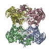 8dheC  8dhlC 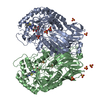 8dhwC 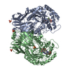 8e72C 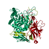 6bjqS S: Starting model for refinement C: citing same article ( |
|---|---|
| Similar structure data |
-Links
 PDBj
PDBj- Assembly
| Deposited unit | 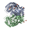
| ||||||||
|---|---|---|---|---|---|---|---|---|---|
| 1 | 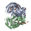
| ||||||||
| Unit cell |
|
-Components
| #1: Protein | Mass: 72233.453 Da / Num. of mol.: 2 Source method: isolated from a genetically manipulated source Source: (gene. exp.) Treponema lecithinolyticum (bacteria) / Production host: #2: Chemical | ChemComp-SO4 /   Mass: 96.063 Da / Num. of mol.: 9 / Source method: obtained synthetically / Formula: SO4 Mass: 96.063 Da / Num. of mol.: 9 / Source method: obtained synthetically / Formula: SO4#3: Chemical | ChemComp-MPD / (   Mass: 118.174 Da / Num. of mol.: 5 / Source method: obtained synthetically / Formula: C6H14O2 / Comment: precipitant*YM Mass: 118.174 Da / Num. of mol.: 5 / Source method: obtained synthetically / Formula: C6H14O2 / Comment: precipitant*YM#4: Chemical | ChemComp-CL /   Mass: 35.453 Da / Num. of mol.: 4 / Source method: obtained synthetically / Formula: Cl Mass: 35.453 Da / Num. of mol.: 4 / Source method: obtained synthetically / Formula: Cl#5: Water | ChemComp-HOH / |  Mass: 18.015 Da / Num. of mol.: 982 / Source method: isolated from a natural source / Formula: H2O Mass: 18.015 Da / Num. of mol.: 982 / Source method: isolated from a natural source / Formula: H2OHas ligand of interest | N | |
|---|
-Experimental details
-Experiment
| Experiment | Method: X-RAY DIFFRACTION / Number of used crystals: 1 |
|---|
- Sample preparation
| Crystal | Density Matthews: 2.26 Å3/Da / Density % sol: 45.6 % |
|---|---|
| Crystal grow | Temperature: 293.15 K / Method: vapor diffusion, hanging drop / pH: 7 / Details: 2 M ammonium sulfate, 0.1 M MOPS, 4% (v/v) MPD |
-Data collection
| Diffraction | Mean temperature: 100 K / Serial crystal experiment: N | ||||||||||||||||||||||||||||||
|---|---|---|---|---|---|---|---|---|---|---|---|---|---|---|---|---|---|---|---|---|---|---|---|---|---|---|---|---|---|---|---|
| Diffraction source | Source: SYNCHROTRON / Site: APS / Beamline: 23-ID-D / Wavelength: 1.03318 Å | ||||||||||||||||||||||||||||||
| Detector | Type: DECTRIS PILATUS3 6M / Detector: PIXEL / Date: Jun 18, 2021 | ||||||||||||||||||||||||||||||
| Radiation | Monochromator: Si(111) / Protocol: SINGLE WAVELENGTH / Monochromatic (M) / Laue (L): M / Scattering type: x-ray | ||||||||||||||||||||||||||||||
| Radiation wavelength | Wavelength: 1.03318 Å / Relative weight: 1 | ||||||||||||||||||||||||||||||
| Reflection | Resolution: 1.6→89.62 Å / Num. obs: 170930 / % possible obs: 99.7 % / Redundancy: 11.1 % / Biso Wilson estimate: 23.66 Å2 / CC1/2: 0.999 / Rmerge(I) obs: 0.092 / Rpim(I) all: 0.028 / Rrim(I) all: 0.097 / Net I/σ(I): 13.1 / Num. measured all: 1896978 / Scaling rejects: 4 | ||||||||||||||||||||||||||||||
| Reflection shell | Diffraction-ID: 1
|
-Phasing
| Phasing | Method: molecular replacement |
|---|
- Processing
| Software |
| |||||||||||||||||||||||||||||||||||||||||||||||||||||||||||||||||||||||||||||||||||||||||||||||||||||||||
|---|---|---|---|---|---|---|---|---|---|---|---|---|---|---|---|---|---|---|---|---|---|---|---|---|---|---|---|---|---|---|---|---|---|---|---|---|---|---|---|---|---|---|---|---|---|---|---|---|---|---|---|---|---|---|---|---|---|---|---|---|---|---|---|---|---|---|---|---|---|---|---|---|---|---|---|---|---|---|---|---|---|---|---|---|---|---|---|---|---|---|---|---|---|---|---|---|---|---|---|---|---|---|---|---|---|---|
| Refinement | Method to determine structure: MOLECULAR REPLACEMENT Starting model: 6BJQ Resolution: 1.6→49.15 Å / SU ML: 0.21 / Cross valid method: THROUGHOUT / σ(F): 1.33 / Phase error: 21.4 / Stereochemistry target values: ML
| |||||||||||||||||||||||||||||||||||||||||||||||||||||||||||||||||||||||||||||||||||||||||||||||||||||||||
| Solvent computation | Shrinkage radii: 0.9 Å / VDW probe radii: 1.11 Å / Solvent model: FLAT BULK SOLVENT MODEL | |||||||||||||||||||||||||||||||||||||||||||||||||||||||||||||||||||||||||||||||||||||||||||||||||||||||||
| Displacement parameters | Biso max: 94.09 Å2 / Biso mean: 29.5718 Å2 / Biso min: 16.35 Å2 | |||||||||||||||||||||||||||||||||||||||||||||||||||||||||||||||||||||||||||||||||||||||||||||||||||||||||
| Refinement step | Cycle: final / Resolution: 1.6→49.15 Å
| |||||||||||||||||||||||||||||||||||||||||||||||||||||||||||||||||||||||||||||||||||||||||||||||||||||||||
| LS refinement shell | Refine-ID: X-RAY DIFFRACTION / Rfactor Rfree error: 0 / Total num. of bins used: 14
| |||||||||||||||||||||||||||||||||||||||||||||||||||||||||||||||||||||||||||||||||||||||||||||||||||||||||
| Refinement TLS params. | Method: refined / Refine-ID: X-RAY DIFFRACTION
| |||||||||||||||||||||||||||||||||||||||||||||||||||||||||||||||||||||||||||||||||||||||||||||||||||||||||
| Refinement TLS group |
|
