 Movie
Movie Controller

[English] 日本語
 Yorodumi
- PDB-8dfv: Structural Basis of MicroRNA Biogenesis by Dicer-1 and Its Partne... -
+ Open data

- Basic information
| Entry | Database: PDB / ID: 8dfv | ||||||||||||
|---|---|---|---|---|---|---|---|---|---|---|---|---|---|
| Title | Structural Basis of MicroRNA Biogenesis by Dicer-1 and Its Partner Protein Loqs-PB - complex IIa | ||||||||||||
 Components Components |
| ||||||||||||
 Keywords Keywords | RNA BINDING PROTEIN/RNA / Dicer / Dcr-1 / Loquacious / Loqs-P / miRNA / RNA BINDING PROTEIN-RNA complex | ||||||||||||
| Function / homology |  Function and homology information Function and homology informationmitotic cell cycle, embryonic / germarium-derived female germ-line cyst formation / germ-line stem cell division / pole cell formation / MicroRNA (miRNA) biogenesis / segment polarity determination / Small interfering RNA (siRNA) biogenesis / germarium-derived oocyte fate determination / PKR-mediated signaling / female germ-line stem cell asymmetric division ...mitotic cell cycle, embryonic / germarium-derived female germ-line cyst formation / germ-line stem cell division / pole cell formation / MicroRNA (miRNA) biogenesis / segment polarity determination / Small interfering RNA (siRNA) biogenesis / germarium-derived oocyte fate determination / PKR-mediated signaling / female germ-line stem cell asymmetric division / regulation of regulatory ncRNA processing / RISC complex binding / dosage compensation by hyperactivation of X chromosome / pre-miRNA binding / germ-line stem cell population maintenance / ribonuclease III / apoptotic DNA fragmentation / deoxyribonuclease I activity / RISC-loading complex / miRNA metabolic process / ribonuclease III activity / RISC complex assembly / regulatory ncRNA-mediated post-transcriptional gene silencing / miRNA processing / pre-miRNA processing / siRNA binding / siRNA processing / RISC complex / dendrite morphogenesis / response to starvation / RNA endonuclease activity / helicase activity / central nervous system development / double-stranded RNA binding / single-stranded RNA binding / RNA binding / ATP binding / metal ion binding / nucleus / cytoplasm / cytosol Similarity search - Function | ||||||||||||
| Biological species |  | ||||||||||||
| Method | ELECTRON MICROSCOPY / single particle reconstruction / cryo EM / Resolution: 3.06 Å | ||||||||||||
 Authors Authors | Jouravleva, K. / Golovenko, D. / Demo, G. / Dutcher, R.C. / Tanaka Hall, T.M. / Zamore, P.D. / Korostelev, A.A. | ||||||||||||
| Funding support |  United States, 3items United States, 3items
| ||||||||||||
 Citation Citation | Journal: Mol Cell / Year: 2022 Title: Structural basis of microRNA biogenesis by Dicer-1 and its partner protein Loqs-PB. Authors: Karina Jouravleva / Dmitrij Golovenko / Gabriel Demo / Robert C Dutcher / Traci M Tanaka Hall / Phillip D Zamore / Andrei A Korostelev /  Abstract: In animals and plants, Dicer enzymes collaborate with double-stranded RNA-binding domain (dsRBD) proteins to convert precursor-microRNAs (pre-miRNAs) into miRNA duplexes. We report six cryo-EM ...In animals and plants, Dicer enzymes collaborate with double-stranded RNA-binding domain (dsRBD) proteins to convert precursor-microRNAs (pre-miRNAs) into miRNA duplexes. We report six cryo-EM structures of Drosophila Dicer-1 that show how Dicer-1 and its partner Loqs‑PB cooperate (1) before binding pre-miRNA, (2) after binding and in a catalytically competent state, (3) after nicking one arm of the pre-miRNA, and (4) following complete dicing and initial product release. Our reconstructions suggest that pre-miRNA binds a rare, open conformation of the Dicer‑1⋅Loqs‑PB heterodimer. The Dicer-1 dsRBD and three Loqs‑PB dsRBDs form a tight belt around the pre-miRNA, distorting the RNA helix to place the scissile phosphodiester bonds in the RNase III active sites. Pre-miRNA cleavage shifts the dsRBDs and partially closes Dicer-1, which may promote product release. Our data suggest a model for how the Dicer‑1⋅Loqs‑PB complex affects a complete cycle of pre-miRNA recognition, stepwise endonuclease cleavage, and product release. | ||||||||||||
| History |
|
- Structure visualization
| Structure viewer | Molecule: MolmilJmol/JSmol |
|---|
- Downloads & links
-Download
| PDBx/mmCIF format | 8dfv.cif.gz | 542.2 KB | Display | PDBx/mmCIF format |
|---|---|---|---|---|
| PDB format | pdb8dfv.ent.gz | 341.8 KB | Display | PDB format |
| PDBx/mmJSON format | 8dfv.json.gz | Tree view | PDBx/mmJSON format | |
| Others |  Other downloads Other downloads |
-Validation report
| Arichive directory | https://data.pdbj.org/pub/pdb/validation_reports/df/8dfvftp://data.pdbj.org/pub/pdb/validation_reports/df/8dfv | HTTPS FTP |
|---|
-Related structure data
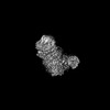
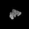
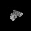
| Related structure data |  27415MC 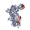 8dg5C 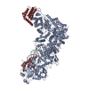 8dg7C 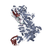 8dgaC 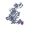 8dgiC 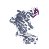 8dgjC M: map data used to model this data C: citing same article ( |
|---|---|
| Similar structure data |


-Links
 PDBj
PDBj


- Assembly
| Deposited unit | 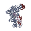
|
|---|---|
| 1 |
|
-Components
| #1: Protein | Mass: 255733.797 Da / Num. of mol.: 1 Source method: isolated from a genetically manipulated source Source: (gene. exp.)  unidentified baculovirus unidentified baculovirusReferences: UniProt: Q9VCU9, Hydrolases; Acting on ester bonds; Endoribonucleases producing 5'-phosphomonoesters | ||
|---|---|---|---|
| #2: RNA chain | Mass: 19102.305 Da / Num. of mol.: 1 / Source method: obtained synthetically / Source: (synth.) | ||
| #3: Protein | Mass: 50149.867 Da / Num. of mol.: 1 Source method: isolated from a genetically manipulated source Source: (gene. exp.) unidentified baculovirus / References: UniProt: Q9VJY9 | ||
| #4: Chemical | ChemComp-CA /   Mass: 40.078 Da / Num. of mol.: 10 / Source method: obtained synthetically / Formula: Ca Mass: 40.078 Da / Num. of mol.: 10 / Source method: obtained synthetically / Formula: CaHas ligand of interest | N | |
-Experimental details
-Experiment
| Experiment | Method: ELECTRON MICROSCOPY |
|---|---|
| EM experiment | Aggregation state: 2D ARRAY / 3D reconstruction method: single particle reconstruction |
- Sample preparation
| Component | Name: Ternary complex of Dicer-1 and Loqs-PB binding pre-miRNA Type: COMPLEX / Entity ID: #1-#3 / Source: MULTIPLE SOURCES |
|---|---|
| Molecular weight | Experimental value: NO |
| Source (natural) | Organism: |
| Source (recombinant) | Organism: unidentified baculovirus |
| Buffer solution | pH: 7.9 |
| Buffer component | Conc.: 20 mM / Name: HEPES-KOH / Formula: HEPES-KOH |
| Specimen | Embedding applied: NO / Shadowing applied: NO / Staining applied: NO / Vitrification applied: YES |
| Specimen support | Grid mesh size: 400 divisions/in. / Grid type: C-flat-1.2/1.3 |
| Vitrification | Instrument: FEI VITROBOT MARK IV / Cryogen name: ETHANE / Humidity: 90 % / Chamber temperature: 277 K |
- Electron microscopy imaging
| Experimental equipment |  Model: Titan Krios / Image courtesy: FEI Company |
|---|---|
| Microscopy | Model: FEI TITAN KRIOS |
| Electron gun | Electron source:  FIELD EMISSION GUN / Accelerating voltage: 300 kV / Illumination mode: FLOOD BEAM FIELD EMISSION GUN / Accelerating voltage: 300 kV / Illumination mode: FLOOD BEAM |
| Electron lens | Mode: BRIGHT FIELD / Nominal defocus max: 3500 nm / Nominal defocus min: 500 nm |
| Image recording | Electron dose: 65.5 e/Å2 / Film or detector model: GATAN K3 (6k x 4k) |
- Processing
| Software |
| ||||||||||||||||||||||||
|---|---|---|---|---|---|---|---|---|---|---|---|---|---|---|---|---|---|---|---|---|---|---|---|---|---|
| EM software |
| ||||||||||||||||||||||||
| CTF correction | Type: NONE | ||||||||||||||||||||||||
| Particle selection | Num. of particles selected: 2023692 | ||||||||||||||||||||||||
| 3D reconstruction | Resolution: 3.06 Å / Resolution method: FSC 0.143 CUT-OFF / Num. of particles: 149443 / Num. of class averages: 1 / Symmetry type: POINT | ||||||||||||||||||||||||
| Refinement | Cross valid method: NONE Stereochemistry target values: GeoStd + Monomer Library + CDL v1.2 | ||||||||||||||||||||||||
| Displacement parameters | Biso mean: 71.84 Å2 | ||||||||||||||||||||||||
| Refine LS restraints |
|
