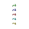
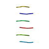
Journal: Chem Sci / Year: 2022 Title: The rippled β-sheet layer configuration-a novel supramolecular architecture based on predictions by Pauling and Corey. Authors: Amaruka Hazari / Michael R Sawaya / Niko Vlahakis / Timothy C Johnstone / David Boyer / Jose Rodriguez / David Eisenberg / Jevgenij A Raskatov / Abstract: The rippled β-sheet is a peptidic structural motif related to but distinct from the pleated β-sheet. Both motifs were predicted in the 1950s by Pauling and Corey. The pleated β-sheet was since ...The rippled β-sheet is a peptidic structural motif related to but distinct from the pleated β-sheet. Both motifs were predicted in the 1950s by Pauling and Corey. The pleated β-sheet was since observed in countless proteins and peptides and is considered common textbook knowledge. Conversely, the rippled β-sheet only gained a meaningful experimental foundation in the past decade, and the first crystal structural study of rippled β-sheets was published as recently as this year. Noteworthy, the crystallized assembly stopped at the rippled β-dimer stage. It did not form the extended, periodic rippled β-sheet layer topography hypothesized by Pauling and Corey, thus calling the validity of their prediction into question. NMR work conducted since moreover shows that certain model peptides rather form pleated and not rippled β-sheets in solution. To determine whether the periodic rippled β-sheet layer configuration is viable, the field urgently needs crystal structures. Here we report on crystal structures of two racemic and one quasi-racemic aggregating peptide systems, all of which yield periodic rippled antiparallel β-sheet layers that are in excellent agreement with the predictions by Pauling and Corey. Our study establishes the rippled β-sheet layer configuration as a motif with general features and opens the road to structure-based design of unique supramolecular architectures.
History
Deposition
Jun 18, 2022
Deposition site: RCSB / Processing site: RCSB
Revision 1.0
Sep 28, 2022
Provider: repository / Type: Initial release
Revision 1.1
May 22, 2024
Group: Data collection / Category: chem_comp_atom / chem_comp_bond
In the structure databanks used in Yorodumi, some data are registered as the other names, "COVID-19 virus" and "2019-nCoV". Here are the details of the virus and the list of structure data.
Jan 31, 2019. EMDB accession codes are about to change! (news from PDBe EMDB page)
EMDB accession codes are about to change! (news from PDBe EMDB page)
The allocation of 4 digits for EMDB accession codes will soon come to an end. Whilst these codes will remain in use, new EMDB accession codes will include an additional digit and will expand incrementally as the available range of codes is exhausted. The current 4-digit format prefixed with “EMD-” (i.e. EMD-XXXX) will advance to a 5-digit format (i.e. EMD-XXXXX), and so on. It is currently estimated that the 4-digit codes will be depleted around Spring 2019, at which point the 5-digit format will come into force.
The EM Navigator/Yorodumi systems omit the EMD- prefix.
Related info.:Q: What is EMD? / ID/Accession-code notation in Yorodumi/EM Navigator
Yorodumi is a browser for structure data from EMDB, PDB, SASBDB, etc.
This page is also the successor to EM Navigator detail page, and also detail information page/front-end page for Omokage search.
The word "yorodu" (or yorozu) is an old Japanese word meaning "ten thousand". "mi" (miru) is to see.
Related info.:EMDB / PDB / SASBDB / Comparison of 3 databanks / Yorodumi Search / Aug 31, 2016. New EM Navigator & Yorodumi / Yorodumi Papers / Jmol/JSmol / Function and homology information / Changes in new EM Navigator and Yorodumi
 Movie
Movie Controller


 Open data
 Basic information
 Components
 Keywords
 X-RAY DIFFRACTION /
  Authors
 Citation
 Structure visualization
 Molmil
 Downloads & links
 Other downloads
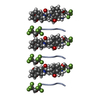
 PDBj
PDBj Assembly
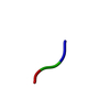
 Mass: 18.015 Da / Num. of mol.: 3 / Source method: isolated from a natural source / Formula: H2O
 Sample preparation
 Processing