 Movie
Movie Controller


+ Open data

- Basic information
| Entry | Database: PDB / ID: 8ddg | ||||||
|---|---|---|---|---|---|---|---|
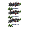
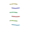
| Title | FYF peptide forms a standard beta-sheet | ||||||
 Components Components | PHE-TYR-PHE | ||||||
 Keywords Keywords | PROTEIN FIBRIL / Rippled beta-sheet / Racemic peptide | ||||||
| Biological species | synthetic construct (others) | ||||||
| Method | ELECTRON CRYSTALLOGRAPHY / electron crystallography /  FOURIER SYNTHESIS / Resolution: 0.9 Å FOURIER SYNTHESIS / Resolution: 0.9 Å | ||||||
 Authors Authors | Sawaya, M.R. / Hazari, A. / Eisenberg, D.E. / Vlahakis, N.W. | ||||||
| Funding support | 1items
| ||||||
 Citation Citation | Journal: Chem Sci / Year: 2022 Title: The rippled β-sheet layer configuration-a novel supramolecular architecture based on predictions by Pauling and Corey. Authors: Amaruka Hazari / Michael R Sawaya / Niko Vlahakis / Timothy C Johnstone / David Boyer / Jose Rodriguez / David Eisenberg / Jevgenij A Raskatov /  Abstract: The rippled β-sheet is a peptidic structural motif related to but distinct from the pleated β-sheet. Both motifs were predicted in the 1950s by Pauling and Corey. The pleated β-sheet was since ...The rippled β-sheet is a peptidic structural motif related to but distinct from the pleated β-sheet. Both motifs were predicted in the 1950s by Pauling and Corey. The pleated β-sheet was since observed in countless proteins and peptides and is considered common textbook knowledge. Conversely, the rippled β-sheet only gained a meaningful experimental foundation in the past decade, and the first crystal structural study of rippled β-sheets was published as recently as this year. Noteworthy, the crystallized assembly stopped at the rippled β-dimer stage. It did not form the extended, periodic rippled β-sheet layer topography hypothesized by Pauling and Corey, thus calling the validity of their prediction into question. NMR work conducted since moreover shows that certain model peptides rather form pleated and not rippled β-sheets in solution. To determine whether the periodic rippled β-sheet layer configuration is viable, the field urgently needs crystal structures. Here we report on crystal structures of two racemic and one quasi-racemic aggregating peptide systems, all of which yield periodic rippled antiparallel β-sheet layers that are in excellent agreement with the predictions by Pauling and Corey. Our study establishes the rippled β-sheet layer configuration as a motif with general features and opens the road to structure-based design of unique supramolecular architectures. | ||||||
| History |
|
- Structure visualization
| Structure viewer | Molecule:  MolmilJmol/JSmol MolmilJmol/JSmol |
|---|
- Downloads & links
-Download
| PDBx/mmCIF format | 8ddg.cif.gz | 13.2 KB | Display | PDBx/mmCIF format |
|---|---|---|---|---|
| PDB format | pdb8ddg.ent.gz | 5 KB | Display | PDB format |
| PDBx/mmJSON format | 8ddg.json.gz | Tree view | PDBx/mmJSON format | |
| Others |  Other downloads Other downloads |
-Validation report
| Arichive directory | https://data.pdbj.org/pub/pdb/validation_reports/dd/8ddgftp://data.pdbj.org/pub/pdb/validation_reports/dd/8ddg | HTTPS FTP |
|---|
-Related structure data
-Links
 PDBj
PDBj- Assembly
| Deposited unit | 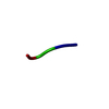
| ||||||||||||
|---|---|---|---|---|---|---|---|---|---|---|---|---|---|
| 1 | 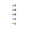
| ||||||||||||
| Unit cell |
|
-Components
| #1: Protein/peptide | Mass: 475.537 Da / Num. of mol.: 1 / Source method: obtained synthetically / Source: (synth.) synthetic construct (others) |
|---|
-Experimental details
-Experiment
| Experiment | Method: ELECTRON CRYSTALLOGRAPHY |
|---|---|
| EM experiment | Aggregation state: 3D ARRAY / 3D reconstruction method: electron crystallography |
- Sample preparation
| Component | Name: FYF peptide / Type: COMPLEX / Entity ID: all / Source: RECOMBINANT |
|---|---|
| Source (natural) | Organism: unidentified (others) |
| Source (recombinant) | Organism: unidentified (others) |
| Buffer solution | pH: 4 |
| Specimen | Embedding applied: NO / Shadowing applied: NO / Staining applied: NO / Vitrification applied: NO / Details: crystal |
-Data collection
| Experimental equipment |  Model: Tecnai F30 / Image courtesy: FEI Company | ||||||||||||||||||||||||||||||||||||||||||||||||||||||||||||||||||||||||||||||||||||||||||||||||||||
|---|---|---|---|---|---|---|---|---|---|---|---|---|---|---|---|---|---|---|---|---|---|---|---|---|---|---|---|---|---|---|---|---|---|---|---|---|---|---|---|---|---|---|---|---|---|---|---|---|---|---|---|---|---|---|---|---|---|---|---|---|---|---|---|---|---|---|---|---|---|---|---|---|---|---|---|---|---|---|---|---|---|---|---|---|---|---|---|---|---|---|---|---|---|---|---|---|---|---|---|---|---|
| Microscopy | Model: FEI TECNAI F30 | ||||||||||||||||||||||||||||||||||||||||||||||||||||||||||||||||||||||||||||||||||||||||||||||||||||
| Electron gun | Electron source: FIELD EMISSION GUN / Accelerating voltage: 300 kV / Illumination mode: FLOOD BEAM | ||||||||||||||||||||||||||||||||||||||||||||||||||||||||||||||||||||||||||||||||||||||||||||||||||||
| Electron lens | Mode: DIFFRACTION / Nominal defocus max: 5000 nm | ||||||||||||||||||||||||||||||||||||||||||||||||||||||||||||||||||||||||||||||||||||||||||||||||||||
| Image recording | Average exposure time: 3 sec. / Electron dose: 0.0192 e/Å2 / Film or detector model: TVIPS TEMCAM-F416 (4k x 4k) | ||||||||||||||||||||||||||||||||||||||||||||||||||||||||||||||||||||||||||||||||||||||||||||||||||||
| EM diffraction | Camera length: 1320 mm | ||||||||||||||||||||||||||||||||||||||||||||||||||||||||||||||||||||||||||||||||||||||||||||||||||||
| EM diffraction shell | Resolution: 0.9→1 Å / Fourier space coverage: 72.2 % / Multiplicity: 4.9 / Num. of structure factors: 332 / Phase residual: 13.5 ° | ||||||||||||||||||||||||||||||||||||||||||||||||||||||||||||||||||||||||||||||||||||||||||||||||||||
| EM diffraction stats | Fourier space coverage: 71 % / High resolution: 0.9 Å / Num. of intensities measured: 5980 / Num. of structure factors: 1237 / Phase error rejection criteria: 20 / Rmerge: 0.113 | ||||||||||||||||||||||||||||||||||||||||||||||||||||||||||||||||||||||||||||||||||||||||||||||||||||
| Diffraction source | Source: ELECTRON MICROSCOPE / Wavelength: 0.0197 Å | ||||||||||||||||||||||||||||||||||||||||||||||||||||||||||||||||||||||||||||||||||||||||||||||||||||
| Detector | Type: TVIPS TEMCAM-F416 / Detector: POSITION SENSITIVE DETECTOR / Date: Dec 14, 2021 | ||||||||||||||||||||||||||||||||||||||||||||||||||||||||||||||||||||||||||||||||||||||||||||||||||||
| Radiation | Protocol: SINGLE WAVELENGTH / Monochromatic (M) / Laue (L): M / Scattering type: electron | ||||||||||||||||||||||||||||||||||||||||||||||||||||||||||||||||||||||||||||||||||||||||||||||||||||
| Radiation wavelength | Wavelength: 0.0197 Å / Relative weight: 1 | ||||||||||||||||||||||||||||||||||||||||||||||||||||||||||||||||||||||||||||||||||||||||||||||||||||
| Reflection | Resolution: 0.9→18.92 Å / Num. obs: 1237 / % possible obs: 71 % / Redundancy: 4.834 % / Biso Wilson estimate: 6.869 Å2 / CC1/2: 0.991 / Rmerge(I) obs: 0.113 / Rrim(I) all: 0.129 / Χ2: 0.932 / Net I/σ(I): 8.63 / Num. measured all: 5980 / Scaling rejects: 11 | ||||||||||||||||||||||||||||||||||||||||||||||||||||||||||||||||||||||||||||||||||||||||||||||||||||
| Reflection shell | Diffraction-ID: 1
|
- Processing
| Software |
| ||||||||||||||||||||||||
|---|---|---|---|---|---|---|---|---|---|---|---|---|---|---|---|---|---|---|---|---|---|---|---|---|---|
| EM 3D crystal entity | ∠α: 90 ° / ∠β: 107.05 ° / ∠γ: 90 ° / A: 23.14 Å / B: 4.84 Å / C: 19.79 Å / Space group name: C2 / Space group num: 5 | ||||||||||||||||||||||||
| CTF correction | Type: NONE | ||||||||||||||||||||||||
| 3D reconstruction | Resolution method: DIFFRACTION PATTERN/LAYERLINES / Symmetry type: 3D CRYSTAL | ||||||||||||||||||||||||
| Atomic model building | Protocol: OTHER / Space: RECIPROCAL | ||||||||||||||||||||||||
| Refinement | Method to determine structure: FOURIER SYNTHESIS / Resolution: 0.9→5.78 Å / SU ML: 0.0753 / Cross valid method: FREE R-VALUE / σ(F): 1.41 / Phase error: 27.6089 Stereochemistry target values: GeoStd + Monomer Library + CDL v1.2
| ||||||||||||||||||||||||
| Solvent computation | Shrinkage radii: 0.9 Å / VDW probe radii: 1.1 Å / Solvent model: FLAT BULK SOLVENT MODEL | ||||||||||||||||||||||||
| Displacement parameters | Biso mean: 3.26 Å2 | ||||||||||||||||||||||||
| Refinement step | Cycle: LAST / Resolution: 0.9→5.78 Å /
| ||||||||||||||||||||||||
| Refine LS restraints |
| ||||||||||||||||||||||||
| LS refinement shell | Resolution: 0.9→5.78 Å
|
