 Movie
Movie Controller


+ Open data

- Basic information
| Entry | Database: PDB / ID: 8dcj | ||||||||||||||||||||||||
|---|---|---|---|---|---|---|---|---|---|---|---|---|---|---|---|---|---|---|---|---|---|---|---|---|---|
| Title | [A:T] Self-Assembled 3D DNA Rhombohedral Tensegrity Triangle | ||||||||||||||||||||||||
 Components Components |
| ||||||||||||||||||||||||
 Keywords Keywords | DNA / tensegrity triangle / synthetic construct / self-assembly | ||||||||||||||||||||||||
| Function / homology | DNA / DNA (> 10) Function and homology information Function and homology information | ||||||||||||||||||||||||
| Biological species | synthetic construct (others) | ||||||||||||||||||||||||
| Method |  X-RAY DIFFRACTION / SYNCHROTRON / MOLECULAR REPLACEMENT / Resolution: 3.33 Å X-RAY DIFFRACTION / SYNCHROTRON / MOLECULAR REPLACEMENT / Resolution: 3.33 Å | ||||||||||||||||||||||||
 Authors Authors | Lu, B. / Vecchioni, S. / Ohayon, Y.P. / Seeman, N.C. / Mao, C. / Sha, R. | ||||||||||||||||||||||||
| Funding support |  United States, 7items United States, 7items
| ||||||||||||||||||||||||
 Citation Citation | Journal: Angew.Chem.Int.Ed.Engl. / Year: 2023 Title: Programmable 3D Hexagonal Geometry of DNA Tensegrity Triangles. Authors: Lu, B. / Woloszyn, K. / Ohayon, Y.P. / Yang, B. / Zhang, C. / Mao, C. / Seeman, N.C. / Vecchioni, S. / Sha, R. | ||||||||||||||||||||||||
| History |
|
- Structure visualization
| Structure viewer | Molecule: MolmilJmol/JSmol |
|---|
- Downloads & links
-Download
| PDBx/mmCIF format | 8dcj.cif.gz | 38 KB | Display | PDBx/mmCIF format |
|---|---|---|---|---|
| PDB format | pdb8dcj.ent.gz | 21.9 KB | Display | PDB format |
| PDBx/mmJSON format | 8dcj.json.gz | Tree view | PDBx/mmJSON format | |
| Others |  Other downloads Other downloads |
-Validation report
| Summary document | 8dcj_validation.pdf.gz | 394.5 KB | Display | wwPDB validaton report |
|---|---|---|---|---|
| Full document | 8dcj_full_validation.pdf.gz | 397.5 KB | Display | |
| Data in XML | 8dcj_validation.xml.gz | 3.7 KB | Display | |
| Data in CIF | 8dcj_validation.cif.gz | 4.4 KB | Display | |
| Arichive directory | https://data.pdbj.org/pub/pdb/validation_reports/dc/8dcjftp://data.pdbj.org/pub/pdb/validation_reports/dc/8dcj | HTTPS FTP |
-Related structure data
| Related structure data | 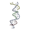 8cs1C 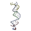 8cs2C 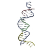 8cs3C 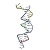 8cs4C 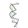 8cs5C 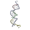 8cs6C 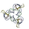 8cs7C 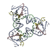 8cs8C 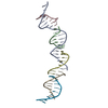 8cymC 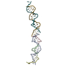 8cynC 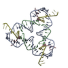 8dagC 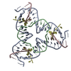 8dahC  5w6wS S: Starting model for refinement C: citing same article ( |
|---|---|
| Similar structure data |
-Links
 PDBj
PDBj


- Assembly
| Deposited unit | 
| ||||||||||||
|---|---|---|---|---|---|---|---|---|---|---|---|---|---|
| 1 | 
| ||||||||||||
| Unit cell |
|
-Components
| #1: DNA chain | Mass: 6441.188 Da / Num. of mol.: 1 / Source method: obtained synthetically / Source: (synth.) synthetic construct (others) |
|---|---|
| #2: DNA chain | Mass: 2082.400 Da / Num. of mol.: 1 / Source method: obtained synthetically / Source: (synth.) synthetic construct (others) |
| #3: DNA chain | Mass: 2129.409 Da / Num. of mol.: 1 / Source method: obtained synthetically / Source: (synth.) synthetic construct (others) |
| #4: DNA chain | Mass: 2143.432 Da / Num. of mol.: 1 / Source method: obtained synthetically / Source: (synth.) synthetic construct (others) |
-Experimental details
-Experiment
| Experiment | Method: X-RAY DIFFRACTION / Number of used crystals: 1 |
|---|
- Sample preparation
| Crystal | Density Matthews: 8.04 Å3/Da / Density % sol: 84.7 % |
|---|---|
| Crystal grow | Temperature: 293 K / Method: vapor diffusion, hanging drop / pH: 9.5 Details: 1.75 M ammonium sulfate, 120 mM Tris, 120 mM Acetic Acid, 6 mM EDTA Temp details: 338-293 at 0.4/hr |
-Data collection
| Diffraction | Mean temperature: 100 K / Serial crystal experiment: N |
|---|---|
| Diffraction source | Source: SYNCHROTRON / Site: APS / Beamline: 17-ID / Wavelength: 1.00743 Å |
| Detector | Type: DECTRIS EIGER2 X 9M / Detector: PIXEL / Date: Feb 21, 2021 |
| Radiation | Protocol: SINGLE WAVELENGTH / Monochromatic (M) / Laue (L): M / Scattering type: x-ray |
| Radiation wavelength | Wavelength: 1.00743 Å / Relative weight: 1 |
| Reflection | Resolution: 3.33→66.2 Å / Num. obs: 3608 / % possible obs: 83.2 % / Redundancy: 10.7 % / Biso Wilson estimate: 172.34 Å2 / CC1/2: 1 / Net I/σ(I): 13.3 |
| Reflection shell | Resolution: 3.33→3.66 Å / Num. unique obs: 194 / CC1/2: 0.34 |
- Processing
| Software |
| ||||||||||||||||||||||||
|---|---|---|---|---|---|---|---|---|---|---|---|---|---|---|---|---|---|---|---|---|---|---|---|---|---|
| Refinement | Method to determine structure: MOLECULAR REPLACEMENT Starting model: 5W6W Resolution: 3.33→33.1 Å / SU ML: 0.3913 / Cross valid method: FREE R-VALUE / σ(F): 1.96 / Phase error: 41.5255 Stereochemistry target values: GeoStd + Monomer Library + CDL v1.2
| ||||||||||||||||||||||||
| Solvent computation | Shrinkage radii: 0.9 Å / VDW probe radii: 1.11 Å / Solvent model: FLAT BULK SOLVENT MODEL | ||||||||||||||||||||||||
| Displacement parameters | Biso mean: 148.31 Å2 | ||||||||||||||||||||||||
| Refinement step | Cycle: LAST / Resolution: 3.33→33.1 Å
| ||||||||||||||||||||||||
| Refine LS restraints |
| ||||||||||||||||||||||||
| LS refinement shell | Resolution: 3.33→33.1 Å
|
