 Movie
Movie Controller

[English] 日本語
 Yorodumi
- PDB-8da1: Crystal structure of Krait alpha-neurotoxin in complex with Centi... -
+ Open data

- Basic information
| Entry | Database: PDB / ID: 8da1 | ||||||||||||
|---|---|---|---|---|---|---|---|---|---|---|---|---|---|
| Title | Crystal structure of Krait alpha-neurotoxin in complex with Centi-LNX-D09 antibody | ||||||||||||
 Components Components |
| ||||||||||||
 Keywords Keywords | IMMUNE SYSTEM / ANTITOXIN/TOXIN / antivenom / 3-finger toxin / alpha-neurotoxin / long neurotoxin 1 / antibody / ANTITOXIN / ANTITOXIN-TOXIN complex | ||||||||||||
| Function / homology |  Function and homology information Function and homology informationacetylcholine receptor inhibitor activity / ion channel regulator activity / toxin activity / extracellular region Similarity search - Function | ||||||||||||
| Biological species |  Homo sapiens (human) Homo sapiens (human) Bungarus multicinctus (many-banded krait) Bungarus multicinctus (many-banded krait) | ||||||||||||
| Method |  X-RAY DIFFRACTION / SYNCHROTRON / MOLECULAR REPLACEMENT / Resolution: 2.67 Å X-RAY DIFFRACTION / SYNCHROTRON / MOLECULAR REPLACEMENT / Resolution: 2.67 Å | ||||||||||||
 Authors Authors | Pletnev, S. / Verardi, R. / Tully, E.S. / Glanville, J. / Kwong, P.D. | ||||||||||||
| Funding support |  United States, 2items United States, 2items
| ||||||||||||
 Citation Citation | Journal: To Be Published Title: Venom protection by antibody from a snakebite hyperimmune subject Authors: Glanville, J. / Andrade, J. / Bellin, M. / Kim, S. / Pletnev, S. / Tsao, D. / Verardi, R. / Bedi, R. / Friede, T. / Tully, E. / Zhang, B. / Bylund, T. / Liu, T. / Kwong, P.D. | ||||||||||||
| History |
|
- Structure visualization
| Structure viewer | Molecule: MolmilJmol/JSmol |
|---|
- Downloads & links
-Download
| PDBx/mmCIF format | 8da1.cif.gz | 112.5 KB | Display | PDBx/mmCIF format |
|---|---|---|---|---|
| PDB format | pdb8da1.ent.gz | 83.4 KB | Display | PDB format |
| PDBx/mmJSON format | 8da1.json.gz | Tree view | PDBx/mmJSON format | |
| Others |  Other downloads Other downloads |
-Validation report
| Arichive directory | https://data.pdbj.org/pub/pdb/validation_reports/da/8da1ftp://data.pdbj.org/pub/pdb/validation_reports/da/8da1 | HTTPS FTP |
|---|
-Related structure data


-Links
 PDBj
PDBj


- Assembly
| Deposited unit | 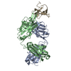
| ||||||||
|---|---|---|---|---|---|---|---|---|---|
| 1 |
| ||||||||
| Unit cell |
|
-Components
| #1: Antibody | Mass: 23339.740 Da / Num. of mol.: 1 Source method: isolated from a genetically manipulated source Source: (gene. exp.) Homo sapiens (human) / Production host: Homo sapiens (human) |
|---|---|
| #2: Antibody | Mass: 25132.959 Da / Num. of mol.: 1 Source method: isolated from a genetically manipulated source Source: (gene. exp.) Homo sapiens (human) / Production host: Homo sapiens (human) |
| #3: Protein | Mass: 8005.281 Da / Num. of mol.: 1 Source method: isolated from a genetically manipulated source Source: (gene. exp.) Bungarus multicinctus (many-banded krait)Production host: Homo sapiens (human) / References: UniProt: P60615 |
| #4: Water | ChemComp-HOH /  Mass: 18.015 Da / Num. of mol.: 3 / Source method: isolated from a natural source / Formula: H2O Mass: 18.015 Da / Num. of mol.: 3 / Source method: isolated from a natural source / Formula: H2O |
| Has protein modification | Y |
-Experimental details
-Experiment
| Experiment | Method: X-RAY DIFFRACTION / Number of used crystals: 1 |
|---|
- Sample preparation
| Crystal | Density Matthews: 3.18 Å3/Da / Density % sol: 61.33 % |
|---|---|
| Crystal grow | Temperature: 293 K / Method: vapor diffusion Details: 40% PEG 400, 5% PEG 3350, 0.1M sodium acetate pH 5.5 |
-Data collection
| Diffraction | Mean temperature: 100 K / Serial crystal experiment: N | |||||||||||||||||||||||||||||||||||||||||||||||||||||||||||||||||||||||||||||||||||||||||||||||||||
|---|---|---|---|---|---|---|---|---|---|---|---|---|---|---|---|---|---|---|---|---|---|---|---|---|---|---|---|---|---|---|---|---|---|---|---|---|---|---|---|---|---|---|---|---|---|---|---|---|---|---|---|---|---|---|---|---|---|---|---|---|---|---|---|---|---|---|---|---|---|---|---|---|---|---|---|---|---|---|---|---|---|---|---|---|---|---|---|---|---|---|---|---|---|---|---|---|---|---|---|---|
| Diffraction source | Source: SYNCHROTRON / Site: APS / Beamline: 22-ID / Wavelength: 1 Å | |||||||||||||||||||||||||||||||||||||||||||||||||||||||||||||||||||||||||||||||||||||||||||||||||||
| Detector | Type: DECTRIS EIGER X 16M / Detector: PIXEL / Date: Oct 10, 2021 | |||||||||||||||||||||||||||||||||||||||||||||||||||||||||||||||||||||||||||||||||||||||||||||||||||
| Radiation | Protocol: SINGLE WAVELENGTH / Monochromatic (M) / Laue (L): M / Scattering type: x-ray | |||||||||||||||||||||||||||||||||||||||||||||||||||||||||||||||||||||||||||||||||||||||||||||||||||
| Radiation wavelength | Wavelength: 1 Å / Relative weight: 1 | |||||||||||||||||||||||||||||||||||||||||||||||||||||||||||||||||||||||||||||||||||||||||||||||||||
| Reflection | Resolution: 2.67→30 Å / Num. obs: 19031 / % possible obs: 98.7 % / Redundancy: 6.7 % / Rmerge(I) obs: 0.248 / Rpim(I) all: 0.103 / Rrim(I) all: 0.27 / Χ2: 1.88 / Net I/σ(I): 7.4 | |||||||||||||||||||||||||||||||||||||||||||||||||||||||||||||||||||||||||||||||||||||||||||||||||||
| Reflection shell | Diffraction-ID: 1
|
- Processing
| Software |
| ||||||||||||||||||||||||||||||||||||||||||||||||||||||||
|---|---|---|---|---|---|---|---|---|---|---|---|---|---|---|---|---|---|---|---|---|---|---|---|---|---|---|---|---|---|---|---|---|---|---|---|---|---|---|---|---|---|---|---|---|---|---|---|---|---|---|---|---|---|---|---|---|---|
| Refinement | Method to determine structure: MOLECULAR REPLACEMENT Starting model: Model found by BALBES software using input sequence Resolution: 2.67→29.51 Å / SU ML: 0.39 / Cross valid method: THROUGHOUT / σ(F): 1.33 / Phase error: 28.54 / Stereochemistry target values: ML
| ||||||||||||||||||||||||||||||||||||||||||||||||||||||||
| Solvent computation | Shrinkage radii: 0.9 Å / VDW probe radii: 1.11 Å / Solvent model: FLAT BULK SOLVENT MODEL | ||||||||||||||||||||||||||||||||||||||||||||||||||||||||
| Refinement step | Cycle: LAST / Resolution: 2.67→29.51 Å
| ||||||||||||||||||||||||||||||||||||||||||||||||||||||||
| Refine LS restraints |
| ||||||||||||||||||||||||||||||||||||||||||||||||||||||||
| LS refinement shell |
|
