 Movie
Movie Controller


+ Open data

- Basic information
| Entry | Database: PDB / ID: 8d9o | ||||||
|---|---|---|---|---|---|---|---|
| Title | De Novo Photosynthetic Reaction Center Protein in Apo-State | ||||||
 Components Components | Reaction Center Maquette | ||||||
 Keywords Keywords | DE NOVO PROTEIN / maquette / protein design / charge separation / artificial photosynthesis | ||||||
| Function / homology | :  Function and homology information Function and homology information | ||||||
| Biological species | synthetic construct (others) | ||||||
| Method |  X-RAY DIFFRACTION / MOLECULAR REPLACEMENT / Resolution: 1.78 Å X-RAY DIFFRACTION / MOLECULAR REPLACEMENT / Resolution: 1.78 Å | ||||||
 Authors Authors | Ennist, N.M. / Stayrook, S.E. / Dutton, P.L. / Moser, C.C. | ||||||
| Funding support |  United States, 1items United States, 1items
| ||||||
 Citation Citation | Journal: Front Mol Biosci / Year: 2022 Title: Rational design of photosynthetic reaction center protein maquettes. Authors: Ennist, N.M. / Stayrook, S.E. / Dutton, P.L. / Moser, C.C. #1: Journal: To Be PublishedTitle: De Novo Protein Design of Photochemical Reaction Centers Authors: Ennist, N.M. / Zhao, Z. / Stayrook, S.E. / Discher, B.M. / Dutton, P.L. / Moser, C.C. | ||||||
| History |
|
- Structure visualization
| Structure viewer | Molecule: MolmilJmol/JSmol |
|---|
- Downloads & links
-Download
| PDBx/mmCIF format | 8d9o.cif.gz | 57.7 KB | Display | PDBx/mmCIF format |
|---|---|---|---|---|
| PDB format | pdb8d9o.ent.gz | 39 KB | Display | PDB format |
| PDBx/mmJSON format | 8d9o.json.gz | Tree view | PDBx/mmJSON format | |
| Others |  Other downloads Other downloads |
-Validation report
| Arichive directory | https://data.pdbj.org/pub/pdb/validation_reports/d9/8d9oftp://data.pdbj.org/pub/pdb/validation_reports/d9/8d9o | HTTPS FTP |
|---|
-Related structure data
| Related structure data |  8d9pC 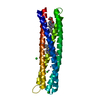 5vjsS S: Starting model for refinement C: citing same article ( |
|---|---|
| Similar structure data |
-Links
 PDBj
PDBj
- Assembly
| Deposited unit | 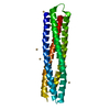
| ||||||||||
|---|---|---|---|---|---|---|---|---|---|---|---|
| 1 |
| ||||||||||
| Unit cell |
| ||||||||||
| Components on special symmetry positions |
|
-Components
| #1: Protein | Mass: 22530.430 Da / Num. of mol.: 1 Source method: isolated from a genetically manipulated source Source: (gene. exp.) synthetic construct (others) / Production host:  | ||||
|---|---|---|---|---|---|
| #2: Chemical | ChemComp-CD /   Mass: 112.411 Da / Num. of mol.: 11 / Source method: obtained synthetically / Formula: Cd / Feature type: SUBJECT OF INVESTIGATION Mass: 112.411 Da / Num. of mol.: 11 / Source method: obtained synthetically / Formula: Cd / Feature type: SUBJECT OF INVESTIGATION#3: Water | ChemComp-HOH / |  Mass: 18.015 Da / Num. of mol.: 95 / Source method: isolated from a natural source / Formula: H2O Mass: 18.015 Da / Num. of mol.: 95 / Source method: isolated from a natural source / Formula: H2OHas ligand of interest | Y | |
-Experimental details
-Experiment
| Experiment | Method: X-RAY DIFFRACTION / Number of used crystals: 1 |
|---|
- Sample preparation
| Crystal | Density Matthews: 1.7 Å3/Da / Density % sol: 27.76 % / Description: Parallelepiped |
|---|---|
| Crystal grow | Temperature: 277 K / Method: vapor diffusion, hanging drop / pH: 4.8 Details: 24% w/v PEG1500, 100 mM CdCl2, 100 mM Na acetate, pH 4.8. Cryoprotectant: 2-methyl-2.4-pentanediol |
-Data collection
| Diffraction | Mean temperature: 100 K / Serial crystal experiment: N |
|---|---|
| Diffraction source | Source: ROTATING ANODE / Type: RIGAKU MICROMAX-007 HF / Wavelength: 1.54178 Å |
| Detector | Type: RIGAKU SATURN 944+ / Detector: CCD / Date: Dec 6, 2013 / Details: Osmic VariMax mirror |
| Radiation | Protocol: SINGLE WAVELENGTH / Monochromatic (M) / Laue (L): M / Scattering type: x-ray |
| Radiation wavelength | Wavelength: 1.54178 Å / Relative weight: 1 |
| Reflection | Resolution: 1.78→18.46 Å / Num. obs: 14128 / % possible obs: 95.1 % / Redundancy: 3.4 % / Biso Wilson estimate: 17.72 Å2 / Rmerge(I) obs: 0.047 / Net I/σ(I): 17.12 |
| Reflection shell | Resolution: 1.783→1.829 Å / Rmerge(I) obs: 0.175 / Mean I/σ(I) obs: 3.79 / Num. unique obs: 737 / CC1/2: 0.95 / % possible all: 68.9 |
- Processing
| Software |
| ||||||||||||||||||||||||||||||||||||||||||
|---|---|---|---|---|---|---|---|---|---|---|---|---|---|---|---|---|---|---|---|---|---|---|---|---|---|---|---|---|---|---|---|---|---|---|---|---|---|---|---|---|---|---|---|
| Refinement | Method to determine structure: MOLECULAR REPLACEMENT Starting model: 5VJS Resolution: 1.78→18.46 Å / SU ML: 0.167 / Cross valid method: FREE R-VALUE / σ(F): 1.36 / Phase error: 20.2235 Stereochemistry target values: GeoStd + Monomer Library + CDL v1.2
| ||||||||||||||||||||||||||||||||||||||||||
| Solvent computation | Shrinkage radii: 0.9 Å / VDW probe radii: 1.11 Å / Solvent model: FLAT BULK SOLVENT MODEL | ||||||||||||||||||||||||||||||||||||||||||
| Displacement parameters | Biso mean: 25.34 Å2 | ||||||||||||||||||||||||||||||||||||||||||
| Refinement step | Cycle: LAST / Resolution: 1.78→18.46 Å
| ||||||||||||||||||||||||||||||||||||||||||
| Refine LS restraints |
| ||||||||||||||||||||||||||||||||||||||||||
| LS refinement shell |
|
