 Movie
Movie Controller

[English] 日本語
 Yorodumi
- PDB-8d87: Fitted crystal structure of the homotrimer of fusion glycoprotein... -
+ Open data

- Basic information
| Entry | Database: PDB / ID: 8d87 | |||||||||
|---|---|---|---|---|---|---|---|---|---|---|
| Title | Fitted crystal structure of the homotrimer of fusion glycoprotein E1 from SFV into subtomogram averaged CHIKV E1 glycoprotein density | |||||||||
 Components Components | Spike glycoprotein E1 | |||||||||
 Keywords Keywords | VIRAL PROTEIN / envelope glycoprotein / membrane fusion / virus | |||||||||
| Function / homology |  Function and homology information Function and homology informationtogavirin / T=4 icosahedral viral capsid / virion assembly / small molecule binding / host cell endoplasmic reticulum / channel activity / host cell endosome / monoatomic ion transmembrane transport / clathrin-dependent endocytosis of virus by host cell / symbiont-mediated suppression of host toll-like receptor signaling pathway ...togavirin / T=4 icosahedral viral capsid / virion assembly / small molecule binding / host cell endoplasmic reticulum / channel activity / host cell endosome / monoatomic ion transmembrane transport / clathrin-dependent endocytosis of virus by host cell / symbiont-mediated suppression of host toll-like receptor signaling pathway / host cell Golgi apparatus / entry receptor-mediated virion attachment to host cell / serine-type endopeptidase activity / viral translational frameshifting / fusion of virus membrane with host endosome membrane / viral envelope / host cell nucleus / host cell plasma membrane / virion membrane / structural molecule activity / proteolysis / RNA binding Similarity search - Function | |||||||||
| Biological species |  Chikungunya virus strain S27-African prototype Chikungunya virus strain S27-African prototype | |||||||||
| Method | ELECTRON MICROSCOPY /  SYNCHROTRON / subtomogram averaging / MAD, MIR / cryo EM / Resolution: 27.2 Å SYNCHROTRON / subtomogram averaging / MAD, MIR / cryo EM / Resolution: 27.2 Å | |||||||||
 Authors Authors | Mangala Prasad, V. / Lee, K.K. | |||||||||
| Funding support |  United States, 2items United States, 2items
| |||||||||
 Citation Citation | Journal: Nat Commun / Year: 2022 Title: Visualization of conformational changes and membrane remodeling leading to genome delivery by viral class-II fusion machinery. Authors: Vidya Mangala Prasad / Jelle S Blijleven / Jolanda M Smit / Kelly K Lee /   Abstract: Chikungunya virus (CHIKV) is a human pathogen that delivers its genome to the host cell cytoplasm through endocytic low pH-activated membrane fusion mediated by class-II fusion proteins. Though ...Chikungunya virus (CHIKV) is a human pathogen that delivers its genome to the host cell cytoplasm through endocytic low pH-activated membrane fusion mediated by class-II fusion proteins. Though structures of prefusion, icosahedral CHIKV are available, structural characterization of virion interaction with membranes has been limited. Here, we have used cryo-electron tomography to visualize CHIKV's complete membrane fusion pathway, identifying key intermediary glycoprotein conformations coupled to membrane remodeling events. Using sub-tomogram averaging, we elucidate features of the low pH-exposed virion, nucleocapsid and full-length E1-glycoprotein's post-fusion structure. Contrary to class-I fusion systems, CHIKV achieves membrane apposition by protrusion of extended E1-glycoprotein homotrimers into the target membrane. The fusion process also features a large hemifusion diaphragm that transitions to a wide pore for intact nucleocapsid delivery. Our analyses provide comprehensive ultrastructural insights into the class-II virus fusion system function and direct mechanistic characterization of the fundamental process of protein-mediated membrane fusion. | |||||||||
| History |
|
- Structure visualization
| Structure viewer | Molecule: MolmilJmol/JSmol |
|---|
- Downloads & links
-Download
| PDBx/mmCIF format | 8d87.cif.gz | 249.9 KB | Display | PDBx/mmCIF format |
|---|---|---|---|---|
| PDB format | pdb8d87.ent.gz | 194.7 KB | Display | PDB format |
| PDBx/mmJSON format | 8d87.json.gz | Tree view | PDBx/mmJSON format | |
| Others |  Other downloads Other downloads |
-Validation report
| Arichive directory | https://data.pdbj.org/pub/pdb/validation_reports/d8/8d87ftp://data.pdbj.org/pub/pdb/validation_reports/d8/8d87 | HTTPS FTP |
|---|
-Related structure data
| Related structure data |  27248MC M: map data used to model this data C: citing same article ( |
|---|---|
| Similar structure data |

-Links
 PDBj
PDBj


- Assembly
| Deposited unit | 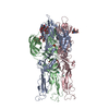
|
|---|---|
| 1 |
|
-Components
-Protein , 1 types, 3 molecules ABC
| #1: Protein | Mass: 42690.125 Da / Num. of mol.: 3 / Fragment: SPIKE GLYCOPROTEIN E1 / Source method: isolated from a natural source Details: The previously determined X-ray crystal structure PDB ID 1RER was rigidly docked into the sub-tomogram averaged map but no further refinement was performed Source: (natural) Chikungunya virus strain S27-African prototypeReferences: UniProt: P03315 |
|---|
-Sugars , 3 types, 3 molecules
| #2: Polysaccharide | 2-acetamido-2-deoxy-beta-D-glucopyranose-(1-2)-beta-D-mannopyranose-(1-3)-beta-D-mannopyranose-(1-4) ...2-acetamido-2-deoxy-beta-D-glucopyranose-(1-2)-beta-D-mannopyranose-(1-3)-beta-D-mannopyranose-(1-4)-2-acetamido-2-deoxy-beta-D-glucopyranose-(1-4)-[beta-L-fucopyranose-(1-6)]2-acetamido-2-deoxy-beta-D-glucopyranose Source method: isolated from a genetically manipulated source |
|---|---|
| #3: Polysaccharide | 2-acetamido-2-deoxy-beta-D-glucopyranose-(1-2)-alpha-D-mannopyranose-(1-3)-[alpha-D-mannopyranose- ...2-acetamido-2-deoxy-beta-D-glucopyranose-(1-2)-alpha-D-mannopyranose-(1-3)-[alpha-D-mannopyranose-(1-6)]beta-D-mannopyranose-(1-4)-2-acetamido-2-deoxy-beta-D-glucopyranose-(1-4)-[alpha-L-fucopyranose-(1-6)]2-acetamido-2-deoxy-alpha-D-glucopyranose Type: oligosaccharide / Mass: 1260.157 Da / Num. of mol.: 1 Source method: isolated from a genetically manipulated source |
| #4: Polysaccharide | 2-acetamido-2-deoxy-beta-D-glucopyranose-(1-4)-2-acetamido-2-deoxy-beta-D-glucopyranose Source method: isolated from a genetically manipulated source |
-Non-polymers , 4 types, 147 molecules 


| #5: Chemical |  Mass: 79.904 Da / Num. of mol.: 3 / Source method: obtained synthetically / Formula: Br Mass: 79.904 Da / Num. of mol.: 3 / Source method: obtained synthetically / Formula: Br#6: Chemical | ChemComp-HO /  Mass: 164.930 Da / Num. of mol.: 4 / Source method: obtained synthetically / Formula: Ho Mass: 164.930 Da / Num. of mol.: 4 / Source method: obtained synthetically / Formula: Ho#7: Chemical | ChemComp-PO4 / |  Mass: 94.971 Da / Num. of mol.: 1 / Source method: obtained synthetically / Formula: PO4 Mass: 94.971 Da / Num. of mol.: 1 / Source method: obtained synthetically / Formula: PO4#8: Water | ChemComp-HOH / | Mass: 18.015 Da / Num. of mol.: 139 / Source method: isolated from a natural source / Formula: H2O |
|---|
-Details
| Has ligand of interest | N |
|---|---|
| Has protein modification | Y |
| Sequence details | The previously determined X-ray crystal structure PDB ID 1RER was rigidly docked into the sub- ...The previously determined X-ray crystal structure PDB ID 1RER was rigidly docked into the sub-tomogram averaged map but no further refinement was performed |
-Experimental details
-Experiment
| Experiment | Method: ELECTRON MICROSCOPY |
|---|---|
| EM experiment | Aggregation state: PARTICLE / 3D reconstruction method: subtomogram averaging |
- Sample preparation
| Component | Name: Sub-tomogram averaged map of post-fusion E1 glycoprotein trimer from Chikungunya virus Type: COMPLEX / Entity ID: #1 / Source: NATURAL |
|---|---|
| Molecular weight | Value: 0.15 MDa / Experimental value: NO |
| Source (natural) | Organism: Chikungunya virus strain S27-African prototype |
| Buffer solution | pH: 5.1 / Details: Hepes Buffer Saline |
| Specimen | Embedding applied: NO / Shadowing applied: NO / Staining applied: NO / Vitrification applied: YES / Details: Sub-volumes picked from liposome membrane surface |
| Specimen support | Grid material: COPPER / Grid mesh size: 400 divisions/in. / Grid type: EMS Lacey Carbon |
| Vitrification | Instrument: FEI VITROBOT MARK IV / Cryogen name: ETHANE / Humidity: 100 % / Chamber temperature: 277 K / Details: blot for 7-8 seconds |
| Crystal | Density Matthews: 5.15 Å3/Da / Density % sol: 76.1 % |
| Crystal grow | pH: 4 Details: PEG 400, NABR, DETERGENT DDAO, HO3+, VAPOR DIFFUSION, HANGING DROP, PH 4, TEMPERATURE 277.0K PH range: 4 |
- Electron microscopy imaging
| Experimental equipment |  Model: Titan Krios / Image courtesy: FEI Company | ||||||||||||||||||||||||
|---|---|---|---|---|---|---|---|---|---|---|---|---|---|---|---|---|---|---|---|---|---|---|---|---|---|
| Microscopy | Model: FEI TITAN KRIOS | ||||||||||||||||||||||||
| Electron gun | Electron source: FIELD EMISSION GUN / Accelerating voltage: 300 kV / Illumination mode: FLOOD BEAM | ||||||||||||||||||||||||
| Electron lens | Mode: BRIGHT FIELD / Nominal magnification: 53000 X / Nominal defocus max: 5000 nm / Nominal defocus min: 2500 nm / Cs: 2.7 mm / C2 aperture diameter: 50 µm / Alignment procedure: COMA FREE | ||||||||||||||||||||||||
| Specimen holder | Cryogen: NITROGEN / Specimen holder model: FEI TITAN KRIOS AUTOGRID HOLDER | ||||||||||||||||||||||||
| Image recording | Electron dose: 70 e/Å2 / Avg electron dose per subtomogram: 70 e/Å2 / Detector mode: SUPER-RESOLUTION / Film or detector model: GATAN K2 SUMMIT (4k x 4k) | ||||||||||||||||||||||||
| Diffraction |
| ||||||||||||||||||||||||
| Diffraction source |
| ||||||||||||||||||||||||
| Detector |
| ||||||||||||||||||||||||
| Radiation |
| ||||||||||||||||||||||||
| Radiation wavelength |
| ||||||||||||||||||||||||
| Reflection | Num. obs: 40912 |


- Processing
| Software |
| |||||||||||||||||||||
|---|---|---|---|---|---|---|---|---|---|---|---|---|---|---|---|---|---|---|---|---|---|---|
| EM software |
| |||||||||||||||||||||
| CTF correction | Type: PHASE FLIPPING AND AMPLITUDE CORRECTION | |||||||||||||||||||||
| Symmetry | Point symmetry: C3 (3 fold cyclic) | |||||||||||||||||||||
| 3D reconstruction | Resolution: 27.2 Å / Resolution method: FSC 0.5 CUT-OFF / Num. of particles: 590 / Symmetry type: POINT | |||||||||||||||||||||
| EM volume selection | Details: low pass filtered map of post-fusion E1--homotrimer crystal structure Num. of tomograms: 40 / Num. of volumes extracted: 591 / Reference model: crystal structure | |||||||||||||||||||||
| Atomic model building | Protocol: RIGID BODY FIT / Space: REAL | |||||||||||||||||||||
| Atomic model building | PDB-ID: 1RER Accession code: 1RER / Source name: PDB / Type: experimental model | |||||||||||||||||||||
| Refinement | Method to determine structure: MAD, MIR / Resolution: 27.2→27.2 Å / σ(F): 2 / Stereochemistry target values: ENGH & HUBER
| |||||||||||||||||||||
| Refinement step | Cycle: LAST / Resolution: 3.2→20 Å
| |||||||||||||||||||||
| LS refinement shell | Highest resolution: 27.2 Å
|

