 Movie
Movie Controller

[English] 日本語
 Yorodumi
- EMDB-27248: Sub-tomogram averaged map of full-length post-fusion CHIKV E1 gly... -
+ Open data

- Basic information
| Entry |  | |||||||||
|---|---|---|---|---|---|---|---|---|---|---|
| Title | Sub-tomogram averaged map of full-length post-fusion CHIKV E1 glycoprotein trimer | |||||||||
 Map data Map data | Sub-tomogram averaged map of membrane attached full-length post-fusion CHIKV E1 glycoprotein trimer | |||||||||
 Sample Sample |
| |||||||||
 Keywords Keywords | envelope glycoprotein / membrane fusion / virus / VIRAL PROTEIN | |||||||||
| Function / homology |  Function and homology information Function and homology informationtogavirin / T=4 icosahedral viral capsid / virion assembly / small molecule binding / host cell endoplasmic reticulum / channel activity / host cell endosome / monoatomic ion transmembrane transport / clathrin-dependent endocytosis of virus by host cell / symbiont-mediated suppression of host toll-like receptor signaling pathway ...togavirin / T=4 icosahedral viral capsid / virion assembly / small molecule binding / host cell endoplasmic reticulum / channel activity / host cell endosome / monoatomic ion transmembrane transport / clathrin-dependent endocytosis of virus by host cell / symbiont-mediated suppression of host toll-like receptor signaling pathway / host cell Golgi apparatus / entry receptor-mediated virion attachment to host cell / serine-type endopeptidase activity / viral translational frameshifting / fusion of virus membrane with host endosome membrane / viral envelope / host cell nucleus / host cell plasma membrane / virion membrane / structural molecule activity / proteolysis / RNA binding Similarity search - Function | |||||||||
| Biological species |  Chikungunya virus strain S27-African prototype Chikungunya virus strain S27-African prototype | |||||||||
| Method | subtomogram averaging / cryo EM / Resolution: 27.2 Å | |||||||||
 Authors Authors | Mangala Prasad V / Lee KK | |||||||||
| Funding support |  United States, 2 items United States, 2 items
| |||||||||
 Citation Citation | Journal: Nat Commun / Year: 2022 Title: Visualization of conformational changes and membrane remodeling leading to genome delivery by viral class-II fusion machinery. Authors: Vidya Mangala Prasad / Jelle S Blijleven / Jolanda M Smit / Kelly K Lee /   Abstract: Chikungunya virus (CHIKV) is a human pathogen that delivers its genome to the host cell cytoplasm through endocytic low pH-activated membrane fusion mediated by class-II fusion proteins. Though ...Chikungunya virus (CHIKV) is a human pathogen that delivers its genome to the host cell cytoplasm through endocytic low pH-activated membrane fusion mediated by class-II fusion proteins. Though structures of prefusion, icosahedral CHIKV are available, structural characterization of virion interaction with membranes has been limited. Here, we have used cryo-electron tomography to visualize CHIKV's complete membrane fusion pathway, identifying key intermediary glycoprotein conformations coupled to membrane remodeling events. Using sub-tomogram averaging, we elucidate features of the low pH-exposed virion, nucleocapsid and full-length E1-glycoprotein's post-fusion structure. Contrary to class-I fusion systems, CHIKV achieves membrane apposition by protrusion of extended E1-glycoprotein homotrimers into the target membrane. The fusion process also features a large hemifusion diaphragm that transitions to a wide pore for intact nucleocapsid delivery. Our analyses provide comprehensive ultrastructural insights into the class-II virus fusion system function and direct mechanistic characterization of the fundamental process of protein-mediated membrane fusion. | |||||||||
| History |
|
- Structure visualization
| Supplemental images |
|---|

- Downloads & links
-EMDB archive
| Map data | emd_27248.map.gz | 232.3 KB | EMDB map data format | |
|---|---|---|---|---|
| Header (meta data) | emd-27248-v30.xmlemd-27248.xml | 18.2 KB 18.2 KB | Display Display | EMDB header |
| FSC (resolution estimation) | emd_27248_fsc.xml | 4.4 KB | Display | FSC data file |
| Images | 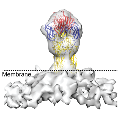 emd_27248.png emd_27248.png | 106.7 KB | ||
| Filedesc metadata | emd-27248.cif.gz | 6.6 KB | ||
| Others | emd_27248_half_map_1.map.gzemd_27248_half_map_2.map.gz | 232.1 KB 232 KB | ||
| Archive directory |  http://ftp.pdbj.org/pub/emdb/structures/EMD-27248ftp://ftp.pdbj.org/pub/emdb/structures/EMD-27248 http://ftp.pdbj.org/pub/emdb/structures/EMD-27248ftp://ftp.pdbj.org/pub/emdb/structures/EMD-27248 | HTTPS FTP |
-Related structure data
| Related structure data |  8d87MC M: atomic model generated by this map C: citing same article ( |
|---|---|
| Similar structure data |

-Links
| EMDB pages | EMDB (EBI/PDBe) / EMDataResource |
|---|---|
| Related items in Molecule of the Month |


-Map
| File | Download / File: emd_27248.map.gz / Format: CCP4 / Size: 251 KB / Type: IMAGE STORED AS FLOATING POINT NUMBER (4 BYTES) | ||||||||||||||||||||||||||||||||||||
|---|---|---|---|---|---|---|---|---|---|---|---|---|---|---|---|---|---|---|---|---|---|---|---|---|---|---|---|---|---|---|---|---|---|---|---|---|---|
| Annotation | Sub-tomogram averaged map of membrane attached full-length post-fusion CHIKV E1 glycoprotein trimer | ||||||||||||||||||||||||||||||||||||
| Projections & slices | Image control
Images are generated by Spider. | ||||||||||||||||||||||||||||||||||||
| Voxel size | X=Y=Z: 6.612 Å | ||||||||||||||||||||||||||||||||||||
| Density |
| ||||||||||||||||||||||||||||||||||||
| Symmetry | Space group: 1 | ||||||||||||||||||||||||||||||||||||
| Details | EMDB XML:
|
 Z (Sec.)
 Y (Row.)
 X (Col.)


-Supplemental data
-Half map: Odd-half map
| File | emd_27248_half_map_1.map | ||||||||||||
|---|---|---|---|---|---|---|---|---|---|---|---|---|---|
| Annotation | Odd-half map | ||||||||||||
| Projections & Slices |
| ||||||||||||
| Density Histograms |


-Half map: Even half map
| File | emd_27248_half_map_2.map | ||||||||||||
|---|---|---|---|---|---|---|---|---|---|---|---|---|---|
| Annotation | Even half map | ||||||||||||
| Projections & Slices |
| ||||||||||||
| Density Histograms |


- Sample components
-Entire : Sub-tomogram averaged map of post-fusion E1 glycoprotein trimer f...
| Entire | Name: Sub-tomogram averaged map of post-fusion E1 glycoprotein trimer from Chikungunya virus |
|---|---|
| Components |
|
-Supramolecule #1: Sub-tomogram averaged map of post-fusion E1 glycoprotein trimer f...
| Supramolecule | Name: Sub-tomogram averaged map of post-fusion E1 glycoprotein trimer from Chikungunya virus type: complex / ID: 1 / Parent: 0 / Macromolecule list: #1 |
|---|---|
| Source (natural) | Organism: Chikungunya virus strain S27-African prototype |
| Molecular weight | Theoretical: 150 KDa |
-Macromolecule #1: Spike glycoprotein E1
| Macromolecule | Name: Spike glycoprotein E1 / type: protein_or_peptide / ID: 1 Details: The previously determined X-ray crystal structure PDB ID 1RER was rigidly docked into the sub-tomogram averaged map but no further refinement was performed Number of copies: 3 / Enantiomer: LEVO |
|---|---|
| Source (natural) | Organism: Chikungunya virus strain S27-African prototype |
| Molecular weight | Theoretical: 42.690125 KDa |
| Sequence | String: YEHSTVMPNV VGFPYKAHIE RPGYSPLTLQ MQVVETSLEP TLNLEYITCE YKTVVPSPYV KCCGASECST KEKPDYQCKV YTGVYPFMW GGAYCFCDSE NTQLSEAYVD RSDVCRHDHA SAYKAHTASL KAKVRVMYGN VNQTVDVYVN GDHAVTIGGT Q FIFGPLSS ...String: YEHSTVMPNV VGFPYKAHIE RPGYSPLTLQ MQVVETSLEP TLNLEYITCE YKTVVPSPYV KCCGASECST KEKPDYQCKV YTGVYPFMW GGAYCFCDSE NTQLSEAYVD RSDVCRHDHA SAYKAHTASL KAKVRVMYGN VNQTVDVYVN GDHAVTIGGT Q FIFGPLSS AWTPFDNKIV VYKDEVFNQD FPPYGSGQPG RFGDIQSRTV ESNDLYANTA LKLARPSPGM VHVPYTQTPS GF KYWLKEK GTALNTKAPF GCQIKTNPVR AMNCAVGNIP VSMNLPDSAF TRIVEAPTII DLTCTVATCT HSSDFGGVLT LTY KTNKNG DCSVHSHSNV ATLQEATAKV KTAGKVTLHF STASASPSFV VSLCSARATC SASCEPPKDH IVPYA UniProtKB: Structural polyprotein |
-Macromolecule #5: BROMIDE ION
| Macromolecule | Name: BROMIDE ION / type: ligand / ID: 5 / Number of copies: 3 / Formula: BR |
|---|---|
| Molecular weight | Theoretical: 79.904 Da |
| Chemical component information |  ChemComp-BR: |
-Macromolecule #6: HOLMIUM ATOM
| Macromolecule | Name: HOLMIUM ATOM / type: ligand / ID: 6 / Number of copies: 4 / Formula: HO |
|---|---|
| Molecular weight | Theoretical: 164.93 Da |
| Chemical component information |  ChemComp-HO: |
-Macromolecule #7: PHOSPHATE ION
| Macromolecule | Name: PHOSPHATE ION / type: ligand / ID: 7 / Number of copies: 1 / Formula: PO4 |
|---|---|
| Molecular weight | Theoretical: 94.971 Da |
| Chemical component information |  ChemComp-PO4: |
-Macromolecule #8: water
| Macromolecule | Name: water / type: ligand / ID: 8 / Number of copies: 139 / Formula: HOH |
|---|---|
| Molecular weight | Theoretical: 18.015 Da |
| Chemical component information |  ChemComp-HOH: |
-Experimental details
-Structure determination
| Method | cryo EM |
|---|---|
 Processing Processing | subtomogram averaging |
| Aggregation state | particle |
-Sample preparation
| Buffer | pH: 5.1 / Details: Hepes Buffer Saline |
|---|---|
| Grid | Model: EMS Lacey Carbon / Material: COPPER / Mesh: 400 / Support film - Material: CARBON / Support film - topology: LACEY / Pretreatment - Type: GLOW DISCHARGE / Pretreatment - Time: 30 sec. / Pretreatment - Atmosphere: AIR |
| Vitrification | Cryogen name: ETHANE / Chamber humidity: 100 % / Chamber temperature: 277 K / Instrument: FEI VITROBOT MARK IV / Details: blot for 7-8 seconds. |
| Details | Sub-volumes picked from liposome membrane surface |
- Electron microscopy
| Microscope | FEI TITAN KRIOS |
|---|---|
| Image recording | Film or detector model: GATAN K2 SUMMIT (4k x 4k) / Detector mode: SUPER-RESOLUTION / Average electron dose: 70.0 e/Å2 |
| Electron beam | Acceleration voltage: 300 kV / Electron source:  FIELD EMISSION GUN FIELD EMISSION GUN |
| Electron optics | C2 aperture diameter: 50.0 µm / Illumination mode: FLOOD BEAM / Imaging mode: BRIGHT FIELD / Cs: 2.7 mm / Nominal defocus max: 5.0 µm / Nominal defocus min: 2.5 µm / Nominal magnification: 53000 |
| Sample stage | Specimen holder model: FEI TITAN KRIOS AUTOGRID HOLDER / Cooling holder cryogen: NITROGEN |
| Experimental equipment |  Model: Titan Krios / Image courtesy: FEI Company |
-Image processing
| Final reconstruction | Applied symmetry - Point group: C3 (3 fold cyclic) / Resolution.type: BY AUTHOR / Resolution: 27.2 Å / Resolution method: FSC 0.5 CUT-OFF / Software - Name: PEET / Number subtomograms used: 590 |
|---|---|
| Extraction | Number tomograms: 40 / Number images used: 591 / Reference model: crystal structure / Software - Name: PEET Details: low pass filtered map of post-fusion E1--homotrimer crystal structure |
| Final angle assignment | Type: NOT APPLICABLE |
| FSC plot (resolution estimation) |  |
-Atomic model buiding 1
| Initial model | PDB ID: Chain - Source name: PDB / Chain - Initial model type: experimental model |
|---|---|
| Refinement | Space: REAL / Protocol: RIGID BODY FIT |
| Output model | PDB-8d87: |

