 Movie
Movie Controller

[English] 日本語
 Yorodumi
- PDB-8d4v: Crystal Structure of Cathepsin G Inhibited by Eap2 from S. aureus -
+ Open data

- Basic information
| Entry | Database: PDB / ID: 8d4v | ||||||
|---|---|---|---|---|---|---|---|
| Title | Crystal Structure of Cathepsin G Inhibited by Eap2 from S. aureus | ||||||
 Components Components |
| ||||||
 Keywords Keywords | HYDROLASE/INHIBITOR / PROTEIN BINDING / Protease Inhibitor / Immune Evasion / Neutrophil / S. aureus / HYDROLASE-INHIBITOR / PROTEIN BINDING complex | ||||||
| Function / homology |  Function and homology information Function and homology informationcathepsin G / biofilm matrix disassembly / neutrophil-mediated killing of gram-positive bacterium / purinergic nucleotide receptor signaling pathway / caspase binding / neutrophil activation / Suppression of apoptosis / Interleukin-1 processing / positive regulation of platelet aggregation / Antimicrobial peptides ...cathepsin G / biofilm matrix disassembly / neutrophil-mediated killing of gram-positive bacterium / purinergic nucleotide receptor signaling pathway / caspase binding / neutrophil activation / Suppression of apoptosis / Interleukin-1 processing / positive regulation of platelet aggregation / Antimicrobial peptides / negative regulation of T cell activation / Activation of Matrix Metalloproteinases / extracellular matrix disassembly / monocyte chemotaxis / defense response to fungus / Metabolism of Angiotensinogen to Angiotensins / Purinergic signaling in leishmaniasis infection / Degradation of the extracellular matrix / angiotensin maturation / secretory granule / serine-type peptidase activity / protein maturation / protein processing / platelet activation / positive regulation of immune response / Regulation of Insulin-like Growth Factor (IGF) transport and uptake by Insulin-like Growth Factor Binding Proteins (IGFBPs) / cytokine-mediated signaling pathway / cytoplasmic stress granule / azurophil granule lumen / peptidase activity / heparin binding / antibacterial humoral response / extracellular matrix / cellular response to lipopolysaccharide / defense response to Gram-negative bacterium / lysosome / defense response to Gram-positive bacterium / immune response / receptor ligand activity / serine-type endopeptidase activity / Neutrophil degranulation / proteolysis / : / extracellular exosome / extracellular region / membrane / nucleus / plasma membrane / cytosol Similarity search - Function | ||||||
| Biological species |  Staphylococcus aureus subsp. aureus Mu50 (bacteria) Staphylococcus aureus subsp. aureus Mu50 (bacteria) Homo sapiens (human) Homo sapiens (human) | ||||||
| Method |  X-RAY DIFFRACTION / SYNCHROTRON / MOLECULAR REPLACEMENT / molecular replacement / Resolution: 1.85 Å X-RAY DIFFRACTION / SYNCHROTRON / MOLECULAR REPLACEMENT / molecular replacement / Resolution: 1.85 Å | ||||||
 Authors Authors | Gido, C.D. / Herdendorf, T.J. / Geisbrecht, B.V. | ||||||
| Funding support |  United States, 1items United States, 1items
| ||||||
 Citation Citation | Journal: J.Biol.Chem. / Year: 2023 Title: Characterization of two distinct neutrophil serine protease-binding modes within a Staphylococcus aureus innate immune evasion protein family. Authors: Gido, C.D. / Herdendorf, T.J. / Geisbrecht, B.V. | ||||||
| History |
|
- Structure visualization
| Structure viewer | Molecule: MolmilJmol/JSmol |
|---|
- Downloads & links
-Download
| PDBx/mmCIF format | 8d4v.cif.gz | 265.3 KB | Display | PDBx/mmCIF format |
|---|---|---|---|---|
| PDB format | pdb8d4v.ent.gz | 213.6 KB | Display | PDB format |
| PDBx/mmJSON format | 8d4v.json.gz | Tree view | PDBx/mmJSON format | |
| Others |  Other downloads Other downloads |
-Validation report
| Arichive directory | https://data.pdbj.org/pub/pdb/validation_reports/d4/8d4vftp://data.pdbj.org/pub/pdb/validation_reports/d4/8d4v | HTTPS FTP |
|---|
-Related structure data
| Related structure data | 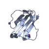 8d4oC  8d4qC  8d4sC  8d4uC 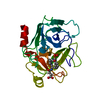 1cghS 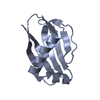 1yn3S C: citing same article ( S: Starting model for refinement |
|---|---|
| Similar structure data |

-Links
 PDBj
PDBj


- Assembly
| Deposited unit | 
| ||||||||
|---|---|---|---|---|---|---|---|---|---|
| 1 | 
| ||||||||
| 2 | 
| ||||||||
| Unit cell |
|
-Components
| #1: Protein | Mass: 25397.131 Da / Num. of mol.: 2 / Source method: isolated from a natural source / Source: (natural) Homo sapiens (human) / Plasmid details: Purified from human sputum / Tissue: Neutrophil / References: UniProt: P08311#2: Protein | Mass: 11023.418 Da / Num. of mol.: 2 Source method: isolated from a genetically manipulated source Source: (gene. exp.) Staphylococcus aureus subsp. aureus Mu50 (bacteria)Strain: Mu50 / ATCC 700699 / Gene: map, SAV1938 / Plasmid: pT7HMT / Production host: #3: Chemical |   Mass: 96.063 Da / Num. of mol.: 2 / Source method: obtained synthetically / Formula: SO4 Mass: 96.063 Da / Num. of mol.: 2 / Source method: obtained synthetically / Formula: SO4#4: Water | ChemComp-HOH / |  Mass: 18.015 Da / Num. of mol.: 224 / Source method: isolated from a natural source / Formula: H2O Mass: 18.015 Da / Num. of mol.: 224 / Source method: isolated from a natural source / Formula: H2OHas ligand of interest | N | Has protein modification | Y | |
|---|
-Experimental details
-Experiment
| Experiment | Method: X-RAY DIFFRACTION / Number of used crystals: 1 |
|---|
- Sample preparation
| Crystal | Density Matthews: 2.13 Å3/Da / Density % sol: 42.3 % |
|---|---|
| Crystal grow | Temperature: 293 K / Method: vapor diffusion, sitting drop / pH: 6.5 Details: 0.1M BisTris (pH 6.5), 0.2M Ammonium Sulfate, 25%(w/v) PEG-3350 |
-Data collection
| Diffraction | Mean temperature: 100 K / Serial crystal experiment: N | |||||||||||||||||||||||||||||||||||||||||||||||||||||||||||||||||||||||||||||||||||||||||||||||||||
|---|---|---|---|---|---|---|---|---|---|---|---|---|---|---|---|---|---|---|---|---|---|---|---|---|---|---|---|---|---|---|---|---|---|---|---|---|---|---|---|---|---|---|---|---|---|---|---|---|---|---|---|---|---|---|---|---|---|---|---|---|---|---|---|---|---|---|---|---|---|---|---|---|---|---|---|---|---|---|---|---|---|---|---|---|---|---|---|---|---|---|---|---|---|---|---|---|---|---|---|---|
| Diffraction source | Source: SYNCHROTRON / Site: APS / Beamline: 22-ID / Wavelength: 1 Å | |||||||||||||||||||||||||||||||||||||||||||||||||||||||||||||||||||||||||||||||||||||||||||||||||||
| Detector | Type: DECTRIS EIGER X 16M / Detector: PIXEL / Date: Nov 4, 2020 | |||||||||||||||||||||||||||||||||||||||||||||||||||||||||||||||||||||||||||||||||||||||||||||||||||
| Radiation | Protocol: SINGLE WAVELENGTH / Monochromatic (M) / Laue (L): M / Scattering type: x-ray | |||||||||||||||||||||||||||||||||||||||||||||||||||||||||||||||||||||||||||||||||||||||||||||||||||
| Radiation wavelength | Wavelength: 1 Å / Relative weight: 1 | |||||||||||||||||||||||||||||||||||||||||||||||||||||||||||||||||||||||||||||||||||||||||||||||||||
| Reflection | Resolution: 1.85→50 Å / Num. obs: 53684 / % possible obs: 99.6 % / Redundancy: 10.1 % / Biso Wilson estimate: 32.64 Å2 / Rmerge(I) obs: 0.242 / Rpim(I) all: 0.075 / Rrim(I) all: 0.253 / Χ2: 1.015 / Net I/σ(I): 7.9 / Num. measured all: 544796 | |||||||||||||||||||||||||||||||||||||||||||||||||||||||||||||||||||||||||||||||||||||||||||||||||||
| Reflection shell | Diffraction-ID: 1
|
-Phasing
| Phasing | Method: molecular replacement |
|---|
- Processing
| Software |
| ||||||||||||||||||||||||||||||||||||||||||||||||||||||||||||||||||||||||||||||||||||||||||||||||||
|---|---|---|---|---|---|---|---|---|---|---|---|---|---|---|---|---|---|---|---|---|---|---|---|---|---|---|---|---|---|---|---|---|---|---|---|---|---|---|---|---|---|---|---|---|---|---|---|---|---|---|---|---|---|---|---|---|---|---|---|---|---|---|---|---|---|---|---|---|---|---|---|---|---|---|---|---|---|---|---|---|---|---|---|---|---|---|---|---|---|---|---|---|---|---|---|---|---|---|---|
| Refinement | Method to determine structure: MOLECULAR REPLACEMENT Starting model: 1CGH, 1YN3 Resolution: 1.85→37.24 Å / SU ML: 0.24 / Cross valid method: THROUGHOUT / σ(F): 0 / Phase error: 32.21 / Stereochemistry target values: ML
| ||||||||||||||||||||||||||||||||||||||||||||||||||||||||||||||||||||||||||||||||||||||||||||||||||
| Solvent computation | Shrinkage radii: 0.9 Å / VDW probe radii: 1.11 Å / Solvent model: FLAT BULK SOLVENT MODEL | ||||||||||||||||||||||||||||||||||||||||||||||||||||||||||||||||||||||||||||||||||||||||||||||||||
| Displacement parameters | Biso max: 118.44 Å2 / Biso mean: 44.2925 Å2 / Biso min: 18.16 Å2 | ||||||||||||||||||||||||||||||||||||||||||||||||||||||||||||||||||||||||||||||||||||||||||||||||||
| Refinement step | Cycle: final / Resolution: 1.85→37.24 Å
| ||||||||||||||||||||||||||||||||||||||||||||||||||||||||||||||||||||||||||||||||||||||||||||||||||
| LS refinement shell | Refine-ID: X-RAY DIFFRACTION / Rfactor Rfree error: 0 / Total num. of bins used: 13
|
