[English] 日本語
Yorodumi
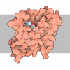
- PDB-8d4t: Mammalian CIV with GDN bound -

+
Open data


ID or keywords:

Loading...

-
Basic information

Entry
Database: PDB / ID: 8d4t
TitleMammalian CIV with GDN bound
Components(Cytochrome c oxidase subunit ...) x 13
KeywordsMEMBRANE PROTEIN / mitochondrial electron transport chain / inhibitor / complex IV
Function / homology
Function and homology information


Complex IV assembly / TP53 Regulates Metabolic Genes / Cytoprotection by HMOX1 / respiratory chain complex IV assembly / mitochondrial respirasome assembly / respiratory chain complex IV / regulation of oxidative phosphorylation / Respiratory electron transport / : / : ...Complex IV assembly / TP53 Regulates Metabolic Genes / Cytoprotection by HMOX1 / respiratory chain complex IV assembly / mitochondrial respirasome assembly / respiratory chain complex IV / regulation of oxidative phosphorylation / Respiratory electron transport / : / : / oxidative phosphorylation / cytochrome-c oxidase / mitochondrial electron transport, cytochrome c to oxygen / cytochrome-c oxidase activity / Mitochondrial protein degradation / electron transport coupled proton transport / ATP synthesis coupled electron transport / enzyme regulator activity / central nervous system development / mitochondrial inner membrane / oxidoreductase activity / copper ion binding / heme binding / mitochondrion / metal ion binding
Similarity search - Function
Cytochrome c oxidase subunit VIIa, metazoa / Cytochrome C oxidase, subunit VIIB / Cytochrome c oxidase subunit IV / Cytochrome C oxidase, subunit VIIB domain superfamily / Mitochondrial cytochrome c oxidase subunit VIc/VIIs / Cytochrome c oxidase, subunit VIIa superfamily / Mitochondrial cytochrome c oxidase subunit VIc/VIIs superfamily / : / Cytochrome c oxidase subunit VIc / Cytochrome C oxidase chain VIIB ...Cytochrome c oxidase subunit VIIa, metazoa / Cytochrome C oxidase, subunit VIIB / Cytochrome c oxidase subunit IV / Cytochrome C oxidase, subunit VIIB domain superfamily / Mitochondrial cytochrome c oxidase subunit VIc/VIIs / Cytochrome c oxidase, subunit VIIa superfamily / Mitochondrial cytochrome c oxidase subunit VIc/VIIs superfamily / : / Cytochrome c oxidase subunit VIc / Cytochrome C oxidase chain VIIB / Cytochrome c oxidase, subunit VIa, conserved site / Cytochrome c oxidase subunit VIa signature. / Cytochrome c oxidase, subunit VIb / : / Cytochrome c oxidase, subunit VIa / Cytochrome c oxidase, subunit VIa superfamily / Cytochrome c oxidase subunit VIa / Cytochrome c oxidase, subunit VIb superfamily / Cytochrome c oxidase subunit 2, C-terminal / Cytochrome oxidase c subunit VIb / Cytochrome c oxidase subunit Vb, zinc binding region signature. / Cytochrome c oxidase subunit VIIc / Cytochrome c oxidase subunit IV family / Cytochrome c oxidase subunit VIIc superfamily / Cytochrome c oxidase subunit IV superfamily / Cytochrome c oxidase subunit VIIc / Cytochrome c oxidase subunit IV / Cytochrome c oxidase, subunit Va/VI / Cytochrome c oxidase, subunit Va/VI superfamily / Cytochrome c oxidase subunit Va / Cytochrome c oxidase subunit VII / Cytochrome c oxidase subunit VII / Cytochrome c oxidase subunit III domain / Cytochrome c oxidase, subunit Vb / Cytochrome c oxidase, subunit Vb superfamily / Cytochrome c oxidase subunit Vb / Cytochrome c oxidase subunit Vb, zinc binding domain profile. / Cytochrome c oxidase, subunit II / Cytochrome c oxidase subunit I domain / Cytochrome C oxidase subunit II, transmembrane domain / Cytochrome C oxidase subunit II, transmembrane domain / Cytochrome oxidase subunit II transmembrane region profile. / Cytochrome c oxidase subunit III / Cytochrome c oxidase subunit III-like / Cytochrome c oxidase, subunit III, 4-helical bundle / Cytochrome c oxidase subunit III / Heme-copper oxidase subunit III family profile. / Cytochrome c oxidase subunit III-like superfamily / Cytochrome c/quinol oxidase subunit II / Copper centre Cu(A) / CO II and nitrous oxide reductase dinuclear copper centers signature. / Cytochrome C oxidase subunit II, transmembrane domain superfamily / Cytochrome c oxidase, subunit I, copper-binding site / Heme-copper oxidase catalytic subunit, copper B binding region signature. / Cytochrome c oxidase-like, subunit I domain / Cytochrome oxidase subunit I profile. / Cytochrome c oxidase subunit I / Cytochrome c oxidase-like, subunit I superfamily / Cytochrome C and Quinol oxidase polypeptide I / Cytochrome C oxidase subunit II, periplasmic domain / Cytochrome c oxidase subunit II-like C-terminal / Cytochrome oxidase subunit II copper A binding domain profile. / Coiled coil-helix-coiled coil-helix (CHCH) domain profile. / Cupredoxin
Similarity search - Domain/homology
COPPER (II) ION / HEME-A / Chem-PEK / Chem-PGV / Cytochrome c oxidase subunit 1 / Cytochrome c oxidase subunit 3 / Cytochrome c oxidase subunit 4 isoform 1, mitochondrial / Cytochrome c oxidase subunit 5A, mitochondrial / Cytochrome c oxidase subunit 5B, mitochondrial / Cytochrome c oxidase subunit 6B1 ...COPPER (II) ION / HEME-A / Chem-PEK / Chem-PGV / Cytochrome c oxidase subunit 1 / Cytochrome c oxidase subunit 3 / Cytochrome c oxidase subunit 4 isoform 1, mitochondrial / Cytochrome c oxidase subunit 5A, mitochondrial / Cytochrome c oxidase subunit 5B, mitochondrial / Cytochrome c oxidase subunit 6B1 / Cytochrome c oxidase subunit 7C, mitochondrial / Cytochrome c oxidase subunit 6C / Cytochrome c oxidase subunit 7A1, mitochondrial / Cytochrome c oxidase subunit 6A2, mitochondrial / Cytochrome c oxidase subunit 7B, mitochondrial / Cytochrome c oxidase subunit 2
Similarity search - Component
Biological speciesBos taurus (cattle)
MethodELECTRON MICROSCOPY / single particle reconstruction / cryo EM / Resolution: 3.1 Å
AuthorsDi Trani, J. / Rubinstein, J.
Funding support Canada, Sweden, 4items
OrganizationGrant numberCountry
Canadian Institutes of Health Research (CIHR)PJT162186 Canada
Knut and Alice Wallenberg Foundation2019.0043 Sweden
Swedish Research Council2018-04619 Sweden
Swedish Research Council016-07213 Sweden
CitationJournal: Proc Natl Acad Sci U S A / Year: 2022
Title: Structural basis of mammalian complex IV inhibition by steroids.
Authors: Justin M Di Trani / Agnes Moe / Daniel Riepl / Patricia Saura / Ville R I Kaila / Peter Brzezinski / John L Rubinstein /
Abstract: The mitochondrial electron transport chain maintains the proton motive force that powers adenosine triphosphate (ATP) synthesis. The energy for this process comes from oxidation of reduced ...The mitochondrial electron transport chain maintains the proton motive force that powers adenosine triphosphate (ATP) synthesis. The energy for this process comes from oxidation of reduced nicotinamide adenine dinucleotide (NADH) and succinate, with the electrons from this oxidation passed via intermediate carriers to oxygen. Complex IV (CIV), the terminal oxidase, transfers electrons from the intermediate electron carrier cytochrome to oxygen, contributing to the proton motive force in the process. Within CIV, protons move through the K and D pathways during turnover. The former is responsible for transferring two protons to the enzyme's catalytic site upon its reduction, where they eventually combine with oxygen and electrons to form water. CIV is the main site for respiratory regulation, and although previous studies showed that steroid binding can regulate CIV activity, little is known about how this regulation occurs. Here, we characterize the interaction between CIV and steroids using a combination of kinetic experiments, structure determination, and molecular simulations. We show that molecules with a sterol moiety, such as glyco-diosgenin and cholesteryl hemisuccinate, reversibly inhibit CIV. Flash photolysis experiments probing the rapid equilibration of electrons within CIV demonstrate that binding of these molecules inhibits proton uptake through the K pathway. Single particle cryogenic electron microscopy (cryo-EM) of CIV with glyco-diosgenin reveals a previously undescribed steroid binding site adjacent to the K pathway, and molecular simulations suggest that the steroid binding modulates the conformational dynamics of key residues and proton transfer kinetics within this pathway. The binding pose of the sterol group sheds light on possible structural gating mechanisms in the CIV catalytic cycle.
History
DepositionJun 2, 2022Deposition site: RCSB / Processing site: RCSB
Revision 1.0Jul 6, 2022Provider: repository / Type: Initial release
Revision 1.1Aug 24, 2022Group: Database references / Category: citation / citation_author
Item: _citation.country / _citation.journal_abbrev ..._citation.country / _citation.journal_abbrev / _citation.journal_id_ASTM / _citation.journal_id_CSD / _citation.journal_id_ISSN / _citation.journal_volume / _citation.page_first / _citation.page_last / _citation.pdbx_database_id_DOI / _citation.pdbx_database_id_PubMed / _citation.title / _citation.year / _citation_author.identifier_ORCID / _citation_author.name

-
Structure visualization

Structure viewerMolecule:
MolmilJmol/JSmol

Downloads & links

-
Assembly

Deposited unit
N: Cytochrome c oxidase subunit 1
O: Cytochrome c oxidase subunit 2
P: Cytochrome c oxidase subunit 3
Q: Cytochrome c oxidase subunit 4 isoform 1, mitochondrial
R: Cytochrome c oxidase subunit 5A, mitochondrial
S: Cytochrome c oxidase subunit 5B, mitochondrial
T: Cytochrome c oxidase subunit 6A2, mitochondrial
U: Cytochrome c oxidase subunit 6B1
V: Cytochrome c oxidase subunit 6C
W: Cytochrome c oxidase subunit 7A1, mitochondrial
X: Cytochrome c oxidase subunit 7B, mitochondrial
Y: Cytochrome c oxidase subunit 7C, mitochondrial
M: Cytochrome c oxidase subunit 8A, mitochondrial
hetero molecules


Theoretical massNumber of molelcules
Total (without water)202,81125
Polymers197,99113
Non-polymers4,82012
Water00
1


  • Idetical with deposited unit
  • defined by author
  • Evidence: scanning transmission electron microscopy
TypeNameSymmetry operationNumber
identity operation1_5551

-
Components

-
Cytochrome c oxidase subunit ... , 13 types, 13 molecules NOPQRSTUVWXYM

#1: Protein Cytochrome c oxidase subunit 1 / Cytochrome c oxidase polypeptide I


Mass: 56964.672 Da / Num. of mol.: 1 / Source method: isolated from a natural source / Source: (natural) Bos taurus (cattle) / References: UniProt: P00396, cytochrome-c oxidase
#2: Protein Cytochrome c oxidase subunit 2 / Cytochrome c oxidase polypeptide II


Mass: 26068.404 Da / Num. of mol.: 1 / Source method: isolated from a natural source / Source: (natural) Bos taurus (cattle) / References: UniProt: P68530
#3: Protein Cytochrome c oxidase subunit 3 / Cytochrome c oxidase polypeptide III


Mass: 29587.180 Da / Num. of mol.: 1 / Source method: isolated from a natural source / Source: (natural) Bos taurus (cattle) / References: UniProt: P00415, cytochrome-c oxidase
#4: Protein Cytochrome c oxidase subunit 4 isoform 1, mitochondrial / Cytochrome c oxidase polypeptide IV / Cytochrome c oxidase subunit IV isoform 1 / COX IV-1


Mass: 16153.474 Da / Num. of mol.: 1 / Source method: isolated from a natural source / Source: (natural) Bos taurus (cattle) / References: UniProt: P00423
#5: Protein Cytochrome c oxidase subunit 5A, mitochondrial / Cytochrome c oxidase polypeptide Va


Mass: 11717.269 Da / Num. of mol.: 1 / Source method: isolated from a natural source / Source: (natural) Bos taurus (cattle) / References: UniProt: P00426
#6: Protein Cytochrome c oxidase subunit 5B, mitochondrial / Cytochrome c oxidase polypeptide VIa / Cytochrome c oxidase polypeptide Vb


Mass: 10018.320 Da / Num. of mol.: 1 / Source method: isolated from a natural source / Source: (natural) Bos taurus (cattle) / References: UniProt: P00428
#7: Protein Cytochrome c oxidase subunit 6A2, mitochondrial / Cytochrome c oxidase polypeptide VIa-heart / COXVIAH / Cytochrome c oxidase polypeptide VIb


Mass: 8368.521 Da / Num. of mol.: 1 / Source method: isolated from a natural source / Source: (natural) Bos taurus (cattle) / References: UniProt: P07471
#8: Protein Cytochrome c oxidase subunit 6B1 / Cytochrome c oxidase polypeptide VII / Cytochrome c oxidase subunit AED / Cytochrome c oxidase ...Cytochrome c oxidase polypeptide VII / Cytochrome c oxidase subunit AED / Cytochrome c oxidase subunit VIb isoform 1 / COX VIb-1


Mass: 9282.420 Da / Num. of mol.: 1 / Source method: isolated from a natural source / Source: (natural) Bos taurus (cattle) / References: UniProt: P00429
#9: Protein Cytochrome c oxidase subunit 6C / Cytochrome c oxidase polypeptide VIc / Cytochrome c oxidase subunit STA


Mass: 8235.723 Da / Num. of mol.: 1 / Source method: isolated from a natural source / Source: (natural) Bos taurus (cattle) / References: UniProt: P04038
#10: Protein Cytochrome c oxidase subunit 7A1, mitochondrial / Cytochrome c oxidase subunit VIIIc / VIIIC / Cytochrome c oxidase subunit VIIa-heart / Cytochrome c ...Cytochrome c oxidase subunit VIIIc / VIIIC / Cytochrome c oxidase subunit VIIa-heart / Cytochrome c oxidase subunit VIIa-H / Cytochrome c oxidase subunit VIIa-muscle / Cytochrome c oxidase subunit VIIa-M


Mass: 6189.105 Da / Num. of mol.: 1 / Source method: isolated from a natural source / Source: (natural) Bos taurus (cattle) / References: UniProt: P07470
#11: Protein/peptide Cytochrome c oxidase subunit 7B, mitochondrial / Cytochrome c oxidase polypeptide VIIb / IHQ


Mass: 5273.975 Da / Num. of mol.: 1 / Source method: isolated from a natural source / Source: (natural) Bos taurus (cattle) / References: UniProt: P13183
#12: Protein/peptide Cytochrome c oxidase subunit 7C, mitochondrial / Cytochrome c oxidase polypeptide VIIIA / Cytochrome c oxidase polypeptide VIIc


Mass: 5362.319 Da / Num. of mol.: 1 / Source method: isolated from a natural source / Source: (natural) Bos taurus (cattle) / References: UniProt: P00430
#13: Protein/peptide Cytochrome c oxidase subunit 8A, mitochondrial


Mass: 4769.651 Da / Num. of mol.: 1 / Source method: isolated from a natural source / Source: (natural) Bos taurus (cattle)

-
Non-polymers , 8 types, 12 molecules
#14: Chemical ChemComp-CU / COPPER (II) ION


Mass: 63.546 Da / Num. of mol.: 3 / Source method: obtained synthetically / Formula: Cu
#15: Chemical ChemComp-MG / MAGNESIUM ION


Mass: 24.305 Da / Num. of mol.: 1 / Source method: obtained synthetically / Formula: Mg
#16: Chemical ChemComp-NA / SODIUM ION


Mass: 22.990 Da / Num. of mol.: 1 / Source method: obtained synthetically / Formula: Na
#17: Chemical ChemComp-HEA / HEME-A


Mass: 852.837 Da / Num. of mol.: 2 / Source method: obtained synthetically / Formula: C49H56FeN4O6
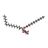
#18: Chemical ChemComp-PGV / (1R)-2-{[{[(2S)-2,3-DIHYDROXYPROPYL]OXY}(HYDROXY)PHOSPHORYL]OXY}-1-[(PALMITOYLOXY)METHYL]ETHYL (11E)-OCTADEC-11-ENOATE / PHOSPHATIDYLGLYCEROL / 2-VACCENOYL-1-PALMITOYL-SN-GLYCEROL-3-PHOSPHOGLYCEROL


Mass: 749.007 Da / Num. of mol.: 2 / Source method: obtained synthetically / Formula: C40H77O10P / Comment: phospholipid*YM
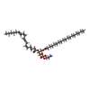
#19: Chemical ChemComp-PEK / (1S)-2-{[(2-AMINOETHOXY)(HYDROXY)PHOSPHORYL]OXY}-1-[(STEAROYLOXY)METHYL]ETHYL (5E,8E,11E,14E)-ICOSA-5,8,11,14-TETRAENOATE / PHOSPHATIDYLETHANOLAMINE / 2-ARACHIDONOYL-1-STEAROYL-SN-GLYCEROL-3-PHOSPHOETHANOLAMINE


Mass: 768.055 Da / Num. of mol.: 1 / Source method: obtained synthetically / Formula: C43H78NO8P / Comment: phospholipid*YM
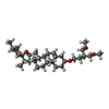
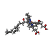
#20: Chemical ChemComp-9Z9 / (3beta,14beta,17beta,25R)-3-[4-methoxy-3-(methoxymethyl)butoxy]spirost-5-en


Mass: 544.805 Da / Num. of mol.: 1 / Source method: obtained synthetically / Formula: C34H56O5 / Feature type: SUBJECT OF INVESTIGATION / Comment: detergent*YM
#21: Chemical ChemComp-ZN / ZINC ION


Mass: 65.409 Da / Num. of mol.: 1 / Source method: obtained synthetically / Formula: Zn

-
Details

Has ligand of interestY

-
Experimental details

-
Experiment

ExperimentMethod: ELECTRON MICROSCOPY
EM experimentAggregation state: PARTICLE / 3D reconstruction method: single particle reconstruction

-
Sample preparation

ComponentName: Cytochrome c oxidase / Type: COMPLEX / Entity ID: #1-#13 / Source: NATURAL
Molecular weightExperimental value: NO
Source (natural)Organism: Bos taurus (cattle)
Buffer solutionpH: 7.5
SpecimenEmbedding applied: NO / Shadowing applied: NO / Staining applied: NO / Vitrification applied: YES
VitrificationCryogen name: ETHANE-PROPANE

-
Electron microscopy imaging

Experimental equipment
Model: Titan Krios / Image courtesy: FEI Company
MicroscopyModel: FEI TITAN KRIOS
Electron gunElectron source: FIELD EMISSION GUN / Accelerating voltage: 300 kV / Illumination mode: FLOOD BEAM
Electron lensMode: BRIGHT FIELD / Nominal defocus max: 3000 nm / Nominal defocus min: 2000 nm
Image recordingElectron dose: 42.7 e/Å2 / Film or detector model: FEI FALCON IV (4k x 4k)

-
Processing

CTF correctionType: PHASE FLIPPING AND AMPLITUDE CORRECTION
3D reconstructionResolution: 3.1 Å / Resolution method: FSC 0.143 CUT-OFF / Num. of particles: 4161 / Symmetry type: POINT
RefinementHighest resolution: 3.1 Å

+
About Yorodumi

-
News

-
Feb 9, 2022. New format data for meta-information of EMDB entries

New format data for meta-information of EMDB entries

  • Version 3 of the EMDB header file is now the official format.
  • The previous official version 1.9 will be removed from the archive.

Related info.:EMDB header

External links:wwPDB to switch to version 3 of the EMDB data model

-
Aug 12, 2020. Covid-19 info

Covid-19 info

URL: https://pdbj.org/emnavi/covid19.php

New page: Covid-19 featured information page in EM Navigator.

Related info.:Covid-19 info / Mar 5, 2020. Novel coronavirus structure data

+
Mar 5, 2020. Novel coronavirus structure data

Novel coronavirus structure data

Related info.:Yorodumi Speices / Aug 12, 2020. Covid-19 info

External links:COVID-19 featured content - PDBj / Molecule of the Month (242):Coronavirus Proteases

+
Jan 31, 2019. EMDB accession codes are about to change! (news from PDBe EMDB page)

EMDB accession codes are about to change! (news from PDBe EMDB page)

  • The allocation of 4 digits for EMDB accession codes will soon come to an end. Whilst these codes will remain in use, new EMDB accession codes will include an additional digit and will expand incrementally as the available range of codes is exhausted. The current 4-digit format prefixed with “EMD-” (i.e. EMD-XXXX) will advance to a 5-digit format (i.e. EMD-XXXXX), and so on. It is currently estimated that the 4-digit codes will be depleted around Spring 2019, at which point the 5-digit format will come into force.
  • The EM Navigator/Yorodumi systems omit the EMD- prefix.

Related info.:Q: What is EMD? / ID/Accession-code notation in Yorodumi/EM Navigator

External links:EMDB Accession Codes are Changing Soon! / Contact to PDBj

+
Jul 12, 2017. Major update of PDB

Major update of PDB

  • wwPDB released updated PDB data conforming to the new PDBx/mmCIF dictionary.
  • This is a major update changing the version number from 4 to 5, and with Remediation, in which all the entries are updated.
  • In this update, many items about electron microscopy experimental information are reorganized (e.g. em_software).
  • Now, EM Navigator and Yorodumi are based on the updated data.

External links:wwPDB Remediation / Enriched Model Files Conforming to OneDep Data Standards Now Available in the PDB FTP Archive

-
Yorodumi

Thousand views of thousand structures

  • Yorodumi is a browser for structure data from EMDB, PDB, SASBDB, etc.
  • This page is also the successor to EM Navigator detail page, and also detail information page/front-end page for Omokage search.
  • The word "yorodu" (or yorozu) is an old Japanese word meaning "ten thousand". "mi" (miru) is to see.

Related info.:EMDB / PDB / SASBDB / Comparison of 3 databanks / Yorodumi Search / Aug 31, 2016. New EM Navigator & Yorodumi / Yorodumi Papers / Jmol/JSmol / Function and homology information / Changes in new EM Navigator and Yorodumi

Read more