 Movie
Movie Controller

[English] 日本語
 Yorodumi
- PDB-8d46: Cryo-electron microscopy structure of human kidney Aldehyde Dehyd... -
+ Open data

- Basic information
| Entry | Database: PDB / ID: 8d46 | ||||||
|---|---|---|---|---|---|---|---|
| Title | Cryo-electron microscopy structure of human kidney Aldehyde Dehydrogenase 1A1 | ||||||
 Components Components | Retinal dehydrogenase 1 | ||||||
 Keywords Keywords | HYDROLASE / aldehyde dehydrogenase / ALDH1A1 | ||||||
| Function / homology |  Function and homology information Function and homology informationfructosamine catabolic process / 3-deoxyglucosone dehydrogenase activity / benzaldehyde dehydrogenase (NAD+) / acetaldehyde dehydrogenase (NAD+) activity / benzaldehyde dehydrogenase (NAD+) activity / aminobutyraldehyde dehydrogenase / retinal dehydrogenase / gamma-aminobutyric acid biosynthetic process / aminobutyraldehyde dehydrogenase (NAD+) activity / maintenance of lens transparency ...fructosamine catabolic process / 3-deoxyglucosone dehydrogenase activity / benzaldehyde dehydrogenase (NAD+) / acetaldehyde dehydrogenase (NAD+) activity / benzaldehyde dehydrogenase (NAD+) activity / aminobutyraldehyde dehydrogenase / retinal dehydrogenase / gamma-aminobutyric acid biosynthetic process / aminobutyraldehyde dehydrogenase (NAD+) activity / maintenance of lens transparency / Fructose catabolism / aldehyde metabolic process / Ethanol oxidation / RA biosynthesis pathway / aldehyde dehydrogenase (NAD+) / androgen binding / cellular detoxification of aldehyde / aldehyde dehydrogenase (NAD+) activity / retinal dehydrogenase (NAD+) activity / retinol metabolic process / negative regulation of cold-induced thermogenesis / retinoid metabolic process / GTPase activator activity / NAD binding / axon / synapse / extracellular exosome / cytoplasm / cytosol Similarity search - Function | ||||||
| Biological species |  Homo sapiens (human) Homo sapiens (human) | ||||||
| Method | ELECTRON MICROSCOPY / single particle reconstruction / cryo EM / Resolution: 2.84 Å | ||||||
 Authors Authors | Lyu, M. / Yu, E.W. | ||||||
| Funding support |  United States, 1items United States, 1items
| ||||||
 Citation Citation | Journal: To Be Published Title: Cryo-electron microscopy structure of human kidney Aldehyde Dehydrogenase 1A1 Authors: Lyu, M. / Yu, E.W. | ||||||
| History |
|
- Structure visualization
| Structure viewer | Molecule: MolmilJmol/JSmol |
|---|
- Downloads & links
-Download
| PDBx/mmCIF format | 8d46.cif.gz | 685.7 KB | Display | PDBx/mmCIF format |
|---|---|---|---|---|
| PDB format | pdb8d46.ent.gz | 584.1 KB | Display | PDB format |
| PDBx/mmJSON format | 8d46.json.gz | Tree view | PDBx/mmJSON format | |
| Others |  Other downloads Other downloads |
-Validation report
| Summary document | 8d46_validation.pdf.gz | 1.2 MB | Display | wwPDB validaton report |
|---|---|---|---|---|
| Full document | 8d46_full_validation.pdf.gz | 1.2 MB | Display | |
| Data in XML | 8d46_validation.xml.gz | 63 KB | Display | |
| Data in CIF | 8d46_validation.cif.gz | 95.2 KB | Display | |
| Arichive directory | https://data.pdbj.org/pub/pdb/validation_reports/d4/8d46ftp://data.pdbj.org/pub/pdb/validation_reports/d4/8d46 | HTTPS FTP |
-Related structure data
| Related structure data |  27176MC M: map data used to model this data C: citing same article ( |
|---|---|
| Similar structure data |
-Links
 PDBj
PDBj


- Assembly
| Deposited unit | 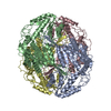
|
|---|---|
| 1 |
|
-Components
| #1: Protein | Mass: 54924.617 Da / Num. of mol.: 4 Source method: isolated from a genetically manipulated source Source: (gene. exp.) Homo sapiens (human) / Gene: ALDH1A1, ALDC, ALDH1, PUMB1 / Production host: Homo sapiens (human)References: UniProt: P00352, Oxidoreductases; Acting on the aldehyde or oxo group of donors; With NAD+ or NADP+ as acceptor, retinal dehydrogenase |
|---|
-Experimental details
-Experiment
| Experiment | Method: ELECTRON MICROSCOPY |
|---|---|
| EM experiment | Aggregation state: PARTICLE / 3D reconstruction method: single particle reconstruction |
- Sample preparation
| Component | Name: Aldehyde dehydrogenase 1A1 / Type: COMPLEX / Entity ID: all / Source: RECOMBINANT |
|---|---|
| Molecular weight | Experimental value: NO |
| Source (natural) | Organism: Homo sapiens (human) |
| Source (recombinant) | Organism: Homo sapiens (human) |
| Buffer solution | pH: 7.5 |
| Specimen | Conc.: 0.7 mg/ml / Embedding applied: NO / Shadowing applied: NO / Staining applied: NO / Vitrification applied: YES |
| Specimen support | Grid material: COPPER / Grid type: Quantifoil R1.2/1.3 |
| Vitrification | Instrument: FEI VITROBOT MARK I / Cryogen name: ETHANE / Humidity: 100 % / Chamber temperature: 277.5 K / Details: blot 15 seconds before plunging |
- Electron microscopy imaging
| Experimental equipment |  Model: Titan Krios / Image courtesy: FEI Company |
|---|---|
| Microscopy | Model: FEI TITAN KRIOS |
| Electron gun | Electron source:  FIELD EMISSION GUN / Accelerating voltage: 300 kV / Illumination mode: OTHER FIELD EMISSION GUN / Accelerating voltage: 300 kV / Illumination mode: OTHER |
| Electron lens | Mode: BRIGHT FIELD / Nominal defocus max: 2500 nm / Nominal defocus min: 1000 nm |
| Image recording | Electron dose: 39 e/Å2 / Film or detector model: GATAN K2 SUMMIT (4k x 4k) |
- Processing
| CTF correction | Type: PHASE FLIPPING AND AMPLITUDE CORRECTION |
|---|---|
| 3D reconstruction | Resolution: 2.84 Å / Resolution method: FSC 0.143 CUT-OFF / Num. of particles: 95743 / Symmetry type: POINT |
| Atomic model building | Protocol: AB INITIO MODEL |
