 Movie
Movie Controller


+ Open data

- Basic information
| Entry | Database: PDB / ID: 8d3a | ||||||
|---|---|---|---|---|---|---|---|
| Title | Crystal Structure of DH475 Fab in complex with Man9 | ||||||
 Components Components |
| ||||||
 Keywords Keywords | IMMUNE SYSTEM/ANTIGEN / FAB FRAGMENT / HIV-1 / ANTIBODY / IMMUNE SYSTEM / IMMUNE SYSTEM-ANTIGEN complex | ||||||
| Function / homology | Immunoglobulins / Immunoglobulin-like / Sandwich / Mainly Beta Function and homology information Function and homology information | ||||||
| Biological species |  Homo sapiens (human) Homo sapiens (human) | ||||||
| Method |  X-RAY DIFFRACTION / SYNCHROTRON / MOLECULAR REPLACEMENT / Resolution: 2.9 Å X-RAY DIFFRACTION / SYNCHROTRON / MOLECULAR REPLACEMENT / Resolution: 2.9 Å | ||||||
 Authors Authors | Finkelstein, M. / Fera, D. | ||||||
| Funding support |  United States, 1items United States, 1items
| ||||||
 Citation Citation | Journal: Front Immunol / Year: 2022 Title: Analysis of two cooperating antibodies unveils immune pressure imposed on HIV Env to elicit a V3-glycan supersite broadly neutralizing antibody lineage. Authors: Maxwell T Finkelstein / Emma Parker Miller / Molly C Erdman / Daniela Fera / Abstract: Elicitation of broadly neutralizing antibodies (bnAbs) is a goal of vaccine design as a strategy for targeting highly divergent strains of HIV-1. Current HIV-1 vaccine design efforts seek to elicit ...Elicitation of broadly neutralizing antibodies (bnAbs) is a goal of vaccine design as a strategy for targeting highly divergent strains of HIV-1. Current HIV-1 vaccine design efforts seek to elicit bnAbs by first eliciting their precursors through prime-boost regimens. This requires an understanding of the co-evolution between viruses and antibodies. Towards this goal, we have analyzed two cooperating antibodies, DH475 and DH272, which exerted pressure on the HIV population in an infected donor, called CH848, to evolve in such a way that it became sensitive to the V3-glycan supersite DH270 bnAb lineage. We obtained a 2.90Å crystal structure of DH475 in complex with the Man glycan and a negative stain EM model of DH272 in complex with the HIV-1 spike trimer, Env. Coupled with additional modeling studies and biochemical data, our studies reveal that DH475 contacts a V3- and V4-glycan dependent epitope accessible on an open or shed Env and that DH272 makes critical contacts with the V1V2 and V3 loops on HIV-1 Env. Using these data, we suggest a prime-boost regimen that may facilitate the initiation of DH270-like bnAb precursors. | ||||||
| History |
|
- Structure visualization
| Structure viewer | Molecule: MolmilJmol/JSmol |
|---|
- Downloads & links
-Download
| PDBx/mmCIF format | 8d3a.cif.gz | 191 KB | Display | PDBx/mmCIF format |
|---|---|---|---|---|
| PDB format | pdb8d3a.ent.gz | 152.3 KB | Display | PDB format |
| PDBx/mmJSON format | 8d3a.json.gz | Tree view | PDBx/mmJSON format | |
| Others |  Other downloads Other downloads |
-Validation report
| Arichive directory | https://data.pdbj.org/pub/pdb/validation_reports/d3/8d3aftp://data.pdbj.org/pub/pdb/validation_reports/d3/8d3a | HTTPS FTP |
|---|
-Related structure data
| Related structure data | 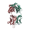 5tppS S: Starting model for refinement C: citing same article ( |
|---|---|
| Similar structure data |
-Links
 PDBj
PDBj


- Assembly
| Deposited unit | 
| ||||||||
|---|---|---|---|---|---|---|---|---|---|
| 1 |
| ||||||||
| Unit cell |
|
-Components
| #1: Antibody | Mass: 24447.354 Da / Num. of mol.: 1 / Fragment: FAB Source method: isolated from a genetically manipulated source Source: (gene. exp.) Homo sapiens (human) / Cell line: HEK293T / Plasmid: pVRC-8400 / Cell line (production host): HEK293T / Production host: Homo sapiens (human) |
|---|---|
| #2: Antibody | Mass: 23166.574 Da / Num. of mol.: 1 Source method: isolated from a genetically manipulated source Source: (gene. exp.) Homo sapiens (human) / Cell line (production host): HEK293T / Production host: Homo sapiens (human) |
| #3: Polysaccharide | alpha-D-mannopyranose-(1-2)-alpha-D-mannopyranose-(1-2)-alpha-D-mannopyranose-(1-3)-[alpha-D- ...alpha-D-mannopyranose-(1-2)-alpha-D-mannopyranose-(1-2)-alpha-D-mannopyranose-(1-3)-[alpha-D-mannopyranose-(1-2)-alpha-D-mannopyranose-(1-3)-[alpha-D-mannopyranose-(1-2)-alpha-D-mannopyranose-(1-6)]alpha-D-mannopyranose-(1-6)]beta-D-mannopyranose-(1-4)-2-acetamido-2-deoxy-beta-D-glucopyranose-(1-4)-2-acetamido-2-deoxy-beta-D-glucopyranose |
| #4: Water | ChemComp-HOH /  Mass: 18.015 Da / Num. of mol.: 65 / Source method: isolated from a natural source / Formula: H2O Mass: 18.015 Da / Num. of mol.: 65 / Source method: isolated from a natural source / Formula: H2O |
| Has ligand of interest | Y |
| Has protein modification | Y |
| Nonpolymer details | The reducing end NAG was altered to place an amine group on C1, which was then reacted with biotin. ...The reducing end NAG was altered to place an amine group on C1, which was then reacted with biotin. The electron density for the biotin, however, is not clear, and so the biotin is not modeled. |
-Experimental details
-Experiment
| Experiment | Method: X-RAY DIFFRACTION / Number of used crystals: 1 |
|---|
- Sample preparation
| Crystal | Density Matthews: 5.25 Å3/Da / Density % sol: 76.59 % |
|---|---|
| Crystal grow | Temperature: 293 K / Method: vapor diffusion, hanging drop / pH: 6 / Details: 1.5 M (NH4)2SO4, 100 mM PIPES, pH 6.0 |
-Data collection
| Diffraction | Mean temperature: 100 K / Serial crystal experiment: N |
|---|---|
| Diffraction source | Source: SYNCHROTRON / Site: APS / Beamline: 24-ID-C / Wavelength: 0.9792 Å |
| Detector | Type: DECTRIS EIGER2 X 16M / Detector: PIXEL / Date: Aug 3, 2016 |
| Radiation | Protocol: SINGLE WAVELENGTH / Monochromatic (M) / Laue (L): M / Scattering type: x-ray |
| Radiation wavelength | Wavelength: 0.9792 Å / Relative weight: 1 |
| Reflection | Resolution: 2.9→76.94 Å / Num. obs: 22928 / % possible obs: 98 % / Redundancy: 3.1 % / CC1/2: 0.993 / Net I/σ(I): 8 |
| Reflection shell | Resolution: 2.9→3.08 Å / Rmerge(I) obs: 1.064 / Num. unique obs: 3672 / % possible all: 99.1 |
- Processing
| Software |
| |||||||||||||||||||||||||||||||||||||||||||||||||||||||||||||||
|---|---|---|---|---|---|---|---|---|---|---|---|---|---|---|---|---|---|---|---|---|---|---|---|---|---|---|---|---|---|---|---|---|---|---|---|---|---|---|---|---|---|---|---|---|---|---|---|---|---|---|---|---|---|---|---|---|---|---|---|---|---|---|---|---|
| Refinement | Method to determine structure: MOLECULAR REPLACEMENT Starting model: 5TPP Resolution: 2.9→76.94 Å / SU ML: 0.45 / Cross valid method: THROUGHOUT / σ(F): 1.34 / Phase error: 28.91 / Stereochemistry target values: ML
| |||||||||||||||||||||||||||||||||||||||||||||||||||||||||||||||
| Solvent computation | Shrinkage radii: 0.9 Å / VDW probe radii: 1.11 Å / Solvent model: FLAT BULK SOLVENT MODEL | |||||||||||||||||||||||||||||||||||||||||||||||||||||||||||||||
| Displacement parameters | Biso max: 146.16 Å2 / Biso mean: 82.3971 Å2 / Biso min: 46.95 Å2 | |||||||||||||||||||||||||||||||||||||||||||||||||||||||||||||||
| Refinement step | Cycle: final / Resolution: 2.9→76.94 Å
| |||||||||||||||||||||||||||||||||||||||||||||||||||||||||||||||
| LS refinement shell | Refine-ID: X-RAY DIFFRACTION / Rfactor Rfree error: 0 / Total num. of bins used: 8
| |||||||||||||||||||||||||||||||||||||||||||||||||||||||||||||||
| Refinement TLS params. | Method: refined / Origin x: -12.7044 Å / Origin y: -45.7812 Å / Origin z: 14.3291 Å
| |||||||||||||||||||||||||||||||||||||||||||||||||||||||||||||||
| Refinement TLS group |
|
