 Movie
Movie Controller


+ Open data

- Basic information
| Entry | Database: PDB / ID: 8d1i | ||||||||||||
|---|---|---|---|---|---|---|---|---|---|---|---|---|---|
| Title | hBest1 1uM Ca2+ (Ca2+-bound) closed state | ||||||||||||
 Components Components | Bestrophin-1 | ||||||||||||
 Keywords Keywords | TRANSPORT PROTEIN / Ion channel / chloride channel / anion channel / pentamer | ||||||||||||
| Function / homology |  Function and homology information Function and homology informationmembrane microdomain / bicarbonate channel activity / gamma-aminobutyric acid secretion, neurotransmission / transepithelial chloride transport / ligand-gated channel activity / intracellularly calcium-gated chloride channel activity / detection of light stimulus involved in visual perception / bicarbonate transmembrane transporter activity / glutamate secretion / chloride transport ...membrane microdomain / bicarbonate channel activity / gamma-aminobutyric acid secretion, neurotransmission / transepithelial chloride transport / ligand-gated channel activity / intracellularly calcium-gated chloride channel activity / detection of light stimulus involved in visual perception / bicarbonate transmembrane transporter activity / glutamate secretion / chloride transport / chloride channel activity / protein complex oligomerization / regulation of calcium ion transport / chloride channel complex / visual perception / basal plasma membrane / regulation of synaptic plasticity / Stimuli-sensing channels / presynapse / monoatomic ion transmembrane transport / basolateral plasma membrane / identical protein binding / membrane / plasma membrane / cytosol Similarity search - Function | ||||||||||||
| Biological species |  Homo sapiens (human) Homo sapiens (human) | ||||||||||||
| Method | ELECTRON MICROSCOPY / single particle reconstruction / cryo EM / Resolution: 1.82 Å | ||||||||||||
 Authors Authors | Owji, A.P. / Kittredge, A. / Hendrickson, W.A. / Tingting, Y. | ||||||||||||
| Funding support |  United States, 3items United States, 3items
| ||||||||||||
 Citation Citation | Journal: Nat Commun / Year: 2022 Title: Structures and gating mechanisms of human bestrophin anion channels. Authors: Aaron P Owji / Jiali Wang / Alec Kittredge / Zada Clark / Yu Zhang / Wayne A Hendrickson / Tingting Yang / Abstract: Bestrophin-1 (Best1) and bestrophin-2 (Best2) are two members of the bestrophin family of calcium (Ca)-activated chloride (Cl) channels with critical involvement in ocular physiology and direct ...Bestrophin-1 (Best1) and bestrophin-2 (Best2) are two members of the bestrophin family of calcium (Ca)-activated chloride (Cl) channels with critical involvement in ocular physiology and direct pathological relevance. Here, we report cryo-EM structures of wild-type human Best1 and Best2 in various states at up to 1.8 Å resolution. Ca-bound Best1 structures illustrate partially open conformations at the two Ca-dependent gates of the channels, in contrast to the fully open conformations observed in Ca-bound Best2, which is in accord with the significantly smaller currents conducted by Best1 in electrophysiological recordings. Comparison of the closed and open states reveals a C-terminal auto-inhibitory segment (AS), which constricts the channel concentrically by wrapping around the channel periphery in an inter-protomer manner and must be released to allow channel opening. Our results demonstrate that removing the AS from Best1 and Best2 results in truncation mutants with similar activities, while swapping the AS between Best1 and Best2 results in chimeric mutants with swapped activities, underlying a key role of the AS in determining paralog specificity among bestrophins. | ||||||||||||
| History |
|
- Structure visualization
| Structure viewer | Molecule: MolmilJmol/JSmol |
|---|
- Downloads & links
-Download
| PDBx/mmCIF format | 8d1i.cif.gz | 435.1 KB | Display | PDBx/mmCIF format |
|---|---|---|---|---|
| PDB format | pdb8d1i.ent.gz | 347.5 KB | Display | PDB format |
| PDBx/mmJSON format | 8d1i.json.gz | Tree view | PDBx/mmJSON format | |
| Others |  Other downloads Other downloads |
-Validation report
| Summary document | 8d1i_validation.pdf.gz | 2.1 MB | Display | wwPDB validaton report |
|---|---|---|---|---|
| Full document | 8d1i_full_validation.pdf.gz | 2.1 MB | Display | |
| Data in XML | 8d1i_validation.xml.gz | 70 KB | Display | |
| Data in CIF | 8d1i_validation.cif.gz | 99.1 KB | Display | |
| Arichive directory | https://data.pdbj.org/pub/pdb/validation_reports/d1/8d1iftp://data.pdbj.org/pub/pdb/validation_reports/d1/8d1i | HTTPS FTP |
-Related structure data
| Related structure data | 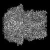 27131MC 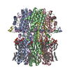 8d1eC 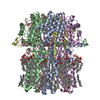 8d1fC  8d1gC 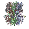 8d1hC 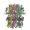 8d1jC 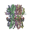 8d1kC 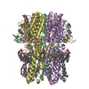 8d1lC 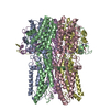 8d1mC 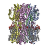 8d1nC 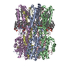 8d1oC M: map data used to model this data C: citing same article ( |
|---|---|
| Similar structure data |
-Links
 PDBj
PDBj- Assembly
| Deposited unit | 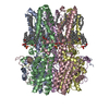
|
|---|---|
| 1 |
|
-Components
| #1: Protein | Mass: 67760.469 Da / Num. of mol.: 5 Source method: isolated from a genetically manipulated source Source: (gene. exp.) Homo sapiens (human) / Gene: BEST1, VMD2 / Cell line (production host): HEK293F / Production host: Homo sapiens (human) / References: UniProt: O76090#2: Chemical | ChemComp-CA /   Mass: 40.078 Da / Num. of mol.: 5 / Source method: obtained synthetically / Formula: Ca / Feature type: SUBJECT OF INVESTIGATION Mass: 40.078 Da / Num. of mol.: 5 / Source method: obtained synthetically / Formula: Ca / Feature type: SUBJECT OF INVESTIGATION#3: Chemical | ChemComp-MC3 /   Mass: 677.933 Da / Num. of mol.: 40 / Source method: obtained synthetically / Formula: C36H72NO8P / Comment: phospholipid*YM Mass: 677.933 Da / Num. of mol.: 40 / Source method: obtained synthetically / Formula: C36H72NO8P / Comment: phospholipid*YM#4: Water | ChemComp-HOH / |  Mass: 18.015 Da / Num. of mol.: 870 / Source method: isolated from a natural source / Formula: H2O Mass: 18.015 Da / Num. of mol.: 870 / Source method: isolated from a natural source / Formula: H2OHas ligand of interest | Y | |
|---|
-Experimental details
-Experiment
| Experiment | Method: ELECTRON MICROSCOPY |
|---|---|
| EM experiment | Aggregation state: PARTICLE / 3D reconstruction method: single particle reconstruction |
- Sample preparation
| Component | Name: hBest1 1uM Ca2+ (Ca2+-bound) closed state / Type: COMPLEX / Entity ID: #1 / Source: RECOMBINANT | ||||||||||||||||||||
|---|---|---|---|---|---|---|---|---|---|---|---|---|---|---|---|---|---|---|---|---|---|
| Molecular weight | Value: 0.338 MDa / Experimental value: NO | ||||||||||||||||||||
| Source (natural) | Organism: Homo sapiens (human) | ||||||||||||||||||||
| Source (recombinant) | Organism: Homo sapiens (human) / Cell: HEK293F | ||||||||||||||||||||
| Buffer solution | pH: 7.8 | ||||||||||||||||||||
| Buffer component |
| ||||||||||||||||||||
| Specimen | Conc.: 5 mg/ml / Embedding applied: NO / Shadowing applied: NO / Staining applied: NO / Vitrification applied: YES | ||||||||||||||||||||
| Vitrification | Instrument: FEI VITROBOT MARK IV / Cryogen name: ETHANE / Humidity: 100 % / Chamber temperature: 283.15 K |
- Electron microscopy imaging
| Experimental equipment |  Model: Titan Krios / Image courtesy: FEI Company |
|---|---|
| Microscopy | Model: TFS KRIOS |
| Electron gun | Electron source:  FIELD EMISSION GUN / Accelerating voltage: 300 kV / Illumination mode: FLOOD BEAM FIELD EMISSION GUN / Accelerating voltage: 300 kV / Illumination mode: FLOOD BEAM |
| Electron lens | Mode: BRIGHT FIELD / Nominal defocus max: 2000 nm / Nominal defocus min: 1000 nm / Cs: 2.7 mm |
| Image recording | Electron dose: 58 e/Å2 / Film or detector model: GATAN K3 BIOQUANTUM (6k x 4k) / Num. of real images: 4670 |
| EM imaging optics | Energyfilter slit width: 20 eV |
- Processing
| EM software |
| ||||||||||||||||||||||||||||||
|---|---|---|---|---|---|---|---|---|---|---|---|---|---|---|---|---|---|---|---|---|---|---|---|---|---|---|---|---|---|---|---|
| CTF correction | Type: NONE | ||||||||||||||||||||||||||||||
| Particle selection | Num. of particles selected: 1727992 | ||||||||||||||||||||||||||||||
| Symmetry | Point symmetry: C5 (5 fold cyclic) | ||||||||||||||||||||||||||||||
| 3D reconstruction | Resolution: 1.82 Å / Resolution method: FSC 0.143 CUT-OFF / Num. of particles: 315602 / Symmetry type: POINT |
