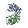
[English] 日本語
Yorodumi
- PDB-8cxl: Structure of NapH3, a vanadium-dependent haloperoxidase homolog c... -

+
Open data


ID or keywords:

Loading...

-
Basic information

Entry
Database: PDB / ID: 8cxl
TitleStructure of NapH3, a vanadium-dependent haloperoxidase homolog catalyzing the stereospecific alpha-hydroxyketone rearrangement reaction in napyradiomycin biosynthesis
ComponentsNapH3
KeywordsBIOSYNTHETIC PROTEIN / Vanadium-dependent haloperoxidase / VHPO / alpha-hydroxyketone rearrangement / napyradiomycin
Biological speciesStreptomyces sp. CNQ-525 (bacteria)
MethodX-RAY DIFFRACTION / SYNCHROTRON / MOLECULAR REPLACEMENT / Resolution: 1.98 Å
AuthorsChen, P.Y.-T. / Chekan, J.R. / Moore, B.S.
Funding support United States, 1items
OrganizationGrant numberCountry
National Institutes of Health/National Institute Of Allergy and Infectious Diseases (NIH/NIAID)R01-AI047818 United States
CitationJournal: Biochemistry / Year: 2022
Title: Structural Basis of Stereospecific Vanadium-Dependent Haloperoxidase Family Enzymes in Napyradiomycin Biosynthesis.
Authors: Chen, P.Y. / Adak, S. / Chekan, J.R. / Liscombe, D.K. / Miyanaga, A. / Bernhardt, P. / Diethelm, S. / Fielding, E.N. / George, J.H. / Miles, Z.D. / Murray, L.A.M. / Steele, T.S. / Winter, J. ...Authors: Chen, P.Y. / Adak, S. / Chekan, J.R. / Liscombe, D.K. / Miyanaga, A. / Bernhardt, P. / Diethelm, S. / Fielding, E.N. / George, J.H. / Miles, Z.D. / Murray, L.A.M. / Steele, T.S. / Winter, J.M. / Noel, J.P. / Moore, B.S.
History
DepositionMay 21, 2022Deposition site: RCSB / Processing site: RCSB
Revision 1.0Aug 31, 2022Provider: repository / Type: Initial release
Revision 1.1Sep 21, 2022Group: Database references / Category: citation / citation_author
Item: _citation.journal_volume / _citation.page_first ..._citation.journal_volume / _citation.page_first / _citation.page_last / _citation_author.identifier_ORCID
Revision 1.2Oct 18, 2023Group: Data collection / Refinement description
Category: chem_comp_atom / chem_comp_bond / pdbx_initial_refinement_model

-
Structure visualization

Structure viewerMolecule:
MolmilJmol/JSmol

Downloads & links

-
Assembly

Deposited unit
A: NapH3
B: NapH3
hetero molecules


Theoretical massNumber of molelcules
Total (without water)103,6105
Polymers103,5262
Non-polymers843
Water9,260514
1


  • Idetical with deposited unit
  • defined by author&software
  • Evidence: gel filtration
TypeNameSymmetry operationNumber
identity operation1_555x,y,z1
Buried area4540 Å2
ΔGint-27 kcal/mol
Surface area32580 Å2
MethodPISA
Unit cell
Length a, b, c (Å)155.447, 96.095, 72.280
Angle α, β, γ (deg.)90.000, 90.000, 90.000
Int Tables number18
Space group name H-MP21212
Space group name HallP22ab
Symmetry operation#1: x,y,z
#2: x+1/2,-y+1/2,-z
#3: -x+1/2,y+1/2,-z
#4: -x,-y,z
Noncrystallographic symmetry (NCS)NCS domain:
IDEns-IDDetails
d_1ens_1(chain "A" and (resid 11 through 26 or resid 29...
d_2ens_1(chain "B" and (resid 11 through 48 or resid 50...

NCS domain segments:
Dom-IDComponent-IDEns-IDBeg label comp-IDEnd label comp-IDLabel asym-IDLabel seq-ID
d_11ens_1PROTHRA7 - 22
d_12ens_1GLYTRPA25 - 44
d_13ens_1THRGLYA48 - 163
d_14ens_1PROTYRA167 - 174
d_15ens_1PROTYRA178 - 230
d_16ens_1ASPALAA232 - 292
d_17ens_1ALAVALA296 - 338
d_18ens_1HISVALA342 - 482
d_21ens_1PROTRPB1 - 36
d_22ens_1THRGLYB40 - 155
d_23ens_1PROTYRB159 - 166
d_24ens_1PROTYRB168 - 220
d_25ens_1ASPALAB224 - 284
d_26ens_1ALAVALB288 - 330
d_27ens_1HISVALB334 - 474

NCS oper: (Code: givenMatrix: (-0.78868324926, 0.567624344346, -0.2361807275), (0.567478778047, 0.524322176392, -0.63486541236), (-0.236530270374, -0.634735266902, -0.735638887055)Vector: -46. ...NCS oper: (Code: given
Matrix: (-0.78868324926, 0.567624344346, -0.2361807275), (0.567478778047, 0.524322176392, -0.63486541236), (-0.236530270374, -0.634735266902, -0.735638887055)
Vector: -46.0964248139, 13.4344949177, -9.12987406766)

-
Components

#1: Protein NapH3


Mass: 51762.980 Da / Num. of mol.: 2
Source method: isolated from a genetically manipulated source
Source: (gene. exp.) Streptomyces sp. CNQ-525 (bacteria) / Gene: napH3 / Production host: Escherichia coli (E. coli)
#2: Chemical ChemComp-MG / MAGNESIUM ION


Mass: 24.305 Da / Num. of mol.: 2 / Source method: obtained synthetically / Formula: Mg
#3: Chemical ChemComp-CL / CHLORIDE ION


Mass: 35.453 Da / Num. of mol.: 1 / Source method: obtained synthetically / Formula: Cl
#4: Water ChemComp-HOH / water


Mass: 18.015 Da / Num. of mol.: 514 / Source method: isolated from a natural source / Formula: H2O
Has ligand of interestY

-
Experimental details

-
Experiment

ExperimentMethod: X-RAY DIFFRACTION / Number of used crystals: 1

-
Sample preparation

CrystalDensity Matthews: 2.61 Å3/Da / Density % sol: 52.82 %
Crystal growTemperature: 279 K / Method: vapor diffusion, hanging drop
Details: 0.1 M bis(2-hydroxyethyl)amino-tris(hydroxymethyl)methane (Bis-Tris) pH 6.5, 0.2 M magnesium chloride , 17% (w/v) PEG 8000

-
Data collection

DiffractionMean temperature: 100 K / Serial crystal experiment: N
Diffraction sourceSource: SYNCHROTRON / Site: ALS / Beamline: 8.2.1 / Wavelength: 0.999912 Å
DetectorType: ADSC QUANTUM 315r / Detector: CCD / Date: Oct 12, 2017
RadiationProtocol: SINGLE WAVELENGTH / Monochromatic (M) / Laue (L): M / Scattering type: x-ray
Radiation wavelengthWavelength: 0.999912 Å / Relative weight: 1
ReflectionResolution: 1.98→30 Å / Num. obs: 75852 / % possible obs: 99.8 % / Redundancy: 6.2 % / Biso Wilson estimate: 28.55 Å2 / Rsym value: 0.096 / Net I/σ(I): 14.8
Reflection shellResolution: 1.98→2.1 Å / Num. unique obs: 12034 / CC1/2: 0.782

-
Processing

Software
NameVersionClassification
PHENIX1.18_3845refinement
XDSdata reduction
XDSdata scaling
PHASERphasing
RefinementMethod to determine structure: MOLECULAR REPLACEMENT
Starting model: 3w36
Resolution: 1.98→29.64 Å / SU ML: 0.1973 / Cross valid method: FREE R-VALUE / σ(F): 1.02 / Phase error: 19.1463
Stereochemistry target values: GeoStd + Monomer Library + CDL v1.2
RfactorNum. reflection% reflection
Rfree0.2007 3793 5 %
Rwork0.1826 72049 -
obs0.1835 75842 99.85 %
Solvent computationShrinkage radii: 0.9 Å / VDW probe radii: 1.11 Å / Solvent model: FLAT BULK SOLVENT MODEL
Displacement parametersBiso mean: 28.55 Å2
Refinement stepCycle: LAST / Resolution: 1.98→29.64 Å
ProteinNucleic acidLigandSolventTotal
Num. atoms7171 0 3 514 7688
Refine LS restraints
Refine-IDTypeDev idealNumber
X-RAY DIFFRACTIONf_bond_d0.00337454
X-RAY DIFFRACTIONf_angle_d0.645410205
X-RAY DIFFRACTIONf_chiral_restr0.04291084
X-RAY DIFFRACTIONf_plane_restr0.00471378
X-RAY DIFFRACTIONf_dihedral_angle_d20.69632644
Refine LS restraints NCSType: Torsion NCS / Rms dev position: 0.482251324203 Å
LS refinement shell
Resolution (Å)Rfactor RfreeNum. reflection RfreeRfactor RworkNum. reflection RworkRefine-ID% reflection obs (%)
1.98-2.010.34241380.29442617X-RAY DIFFRACTION97.42
2.01-2.030.25441360.24852599X-RAY DIFFRACTION100
2.03-2.060.2681400.23292651X-RAY DIFFRACTION100
2.06-2.090.24521390.22042624X-RAY DIFFRACTION99.96
2.09-2.120.2151390.21292645X-RAY DIFFRACTION99.96
2.12-2.150.27521370.21032617X-RAY DIFFRACTION100
2.15-2.190.22381410.19992686X-RAY DIFFRACTION100
2.19-2.230.23471390.20862628X-RAY DIFFRACTION100
2.23-2.270.25581380.20592633X-RAY DIFFRACTION100
2.27-2.310.21091420.19532672X-RAY DIFFRACTION100
2.31-2.360.22451370.19752623X-RAY DIFFRACTION99.96
2.36-2.410.22541410.19772662X-RAY DIFFRACTION99.96
2.41-2.470.21261390.1942639X-RAY DIFFRACTION99.96
2.47-2.530.21271390.19232644X-RAY DIFFRACTION99.96
2.53-2.60.19181390.19032661X-RAY DIFFRACTION100
2.6-2.670.23971420.19322675X-RAY DIFFRACTION99.96
2.67-2.760.21861390.19692650X-RAY DIFFRACTION100
2.76-2.860.22331400.19462654X-RAY DIFFRACTION100
2.86-2.970.21671390.20312655X-RAY DIFFRACTION99.96
2.97-3.110.20941430.20622703X-RAY DIFFRACTION99.96
3.11-3.270.23091400.20552666X-RAY DIFFRACTION100
3.27-3.470.20711420.19492689X-RAY DIFFRACTION100
3.47-3.740.20981420.17872695X-RAY DIFFRACTION99.96
3.74-4.120.15931400.15022694X-RAY DIFFRACTION99.82
4.12-4.710.14481450.13952731X-RAY DIFFRACTION99.9
4.71-5.930.15731450.14572762X-RAY DIFFRACTION99.83
5.93-29.640.14931520.14592874X-RAY DIFFRACTION99.47

+
About Yorodumi

-
News

-
Feb 9, 2022. New format data for meta-information of EMDB entries

New format data for meta-information of EMDB entries

  • Version 3 of the EMDB header file is now the official format.
  • The previous official version 1.9 will be removed from the archive.

Related info.:EMDB header

External links:wwPDB to switch to version 3 of the EMDB data model

-
Aug 12, 2020. Covid-19 info

Covid-19 info

URL: https://pdbjlvh1.pdbj.org/emnavi/covid19.php

New page: Covid-19 featured information page in EM Navigator.

Related info.:Covid-19 info / Mar 5, 2020. Novel coronavirus structure data

+
Mar 5, 2020. Novel coronavirus structure data

Novel coronavirus structure data

Related info.:Yorodumi Speices / Aug 12, 2020. Covid-19 info

External links:COVID-19 featured content - PDBj / Molecule of the Month (242):Coronavirus Proteases

+
Jan 31, 2019. EMDB accession codes are about to change! (news from PDBe EMDB page)

EMDB accession codes are about to change! (news from PDBe EMDB page)

  • The allocation of 4 digits for EMDB accession codes will soon come to an end. Whilst these codes will remain in use, new EMDB accession codes will include an additional digit and will expand incrementally as the available range of codes is exhausted. The current 4-digit format prefixed with “EMD-” (i.e. EMD-XXXX) will advance to a 5-digit format (i.e. EMD-XXXXX), and so on. It is currently estimated that the 4-digit codes will be depleted around Spring 2019, at which point the 5-digit format will come into force.
  • The EM Navigator/Yorodumi systems omit the EMD- prefix.

Related info.:Q: What is EMD? / ID/Accession-code notation in Yorodumi/EM Navigator

External links:EMDB Accession Codes are Changing Soon! / Contact to PDBj

+
Jul 12, 2017. Major update of PDB

Major update of PDB

  • wwPDB released updated PDB data conforming to the new PDBx/mmCIF dictionary.
  • This is a major update changing the version number from 4 to 5, and with Remediation, in which all the entries are updated.
  • In this update, many items about electron microscopy experimental information are reorganized (e.g. em_software).
  • Now, EM Navigator and Yorodumi are based on the updated data.

External links:wwPDB Remediation / Enriched Model Files Conforming to OneDep Data Standards Now Available in the PDB FTP Archive

-
Yorodumi

Thousand views of thousand structures

  • Yorodumi is a browser for structure data from EMDB, PDB, SASBDB, etc.
  • This page is also the successor to EM Navigator detail page, and also detail information page/front-end page for Omokage search.
  • The word "yorodu" (or yorozu) is an old Japanese word meaning "ten thousand". "mi" (miru) is to see.

Related info.:EMDB / PDB / SASBDB / Comparison of 3 databanks / Yorodumi Search / Aug 31, 2016. New EM Navigator & Yorodumi / Yorodumi Papers / Jmol/JSmol / Function and homology information / Changes in new EM Navigator and Yorodumi

Read more