 Movie
Movie Controller

[English] 日本語
 Yorodumi
- PDB-8cxi: Structures of Zika Virus in Complex with Antibodies Targeting E D... -
+ Open data

- Basic information
| Entry | Database: PDB / ID: 8cxi | ||||||||||||
|---|---|---|---|---|---|---|---|---|---|---|---|---|---|
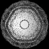
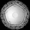
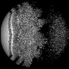
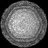
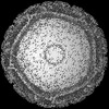
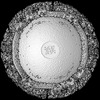
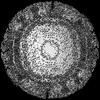
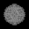
| Title | Structures of Zika Virus in Complex with Antibodies Targeting E Dimer Epitopes and Basis for Neutralization Efficacy | ||||||||||||
 Components Components |
| ||||||||||||
 Keywords Keywords | VIRUS/IMMUNE SYSTEM / ZIKV / scFv antibody / VIRUS-IMMUNE SYSTEM complex | ||||||||||||
| Function / homology |  Function and homology information Function and homology informationlow-density lipoprotein particle receptor binding / regulation of protein-containing complex assembly / symbiont-mediated suppression of host JAK-STAT cascade via inhibition of STAT2 activity / histone deacetylase binding / viral capsid / ribonucleoside triphosphate phosphatase activity / double-stranded RNA binding / regulation of gene expression / molecular adaptor activity / cytoskeleton ...low-density lipoprotein particle receptor binding / regulation of protein-containing complex assembly / symbiont-mediated suppression of host JAK-STAT cascade via inhibition of STAT2 activity / histone deacetylase binding / viral capsid / ribonucleoside triphosphate phosphatase activity / double-stranded RNA binding / regulation of gene expression / molecular adaptor activity / cytoskeleton / methyltransferase cap1 activity / mRNA 5'-cap (guanine-N7-)-methyltransferase activity / RNA helicase activity / protein dimerization activity / symbiont-mediated suppression of host innate immune response / host cell perinuclear region of cytoplasm / host cell endoplasmic reticulum membrane / symbiont-mediated suppression of host type I interferon-mediated signaling pathway / serine-type endopeptidase activity / symbiont-mediated activation of host autophagy / viral RNA genome replication / RNA-directed RNA polymerase activity / fusion of virus membrane with host endosome membrane / symbiont entry into host cell / ubiquitin protein ligase binding / lipid binding / protein kinase binding / virion attachment to host cell / GTP binding / host cell nucleus / virion membrane / structural molecule activity / protein-containing complex / proteolysis / extracellular region / ATP binding / membrane / metal ion binding / nucleus / cytosol Similarity search - Function | ||||||||||||
| Biological species |   Zika virus Zika virus Homo sapiens (human) Homo sapiens (human) | ||||||||||||
| Method | ELECTRON MICROSCOPY / single particle reconstruction / cryo EM / Resolution: 3.4 Å | ||||||||||||
 Authors Authors | Liu, W. / Zhang, X.K. / Gong, D.Y. / Dai, X.H. / Sharma, A. / Zhang, T.H. / Rey, F. / Zhou, Z.H. | ||||||||||||
| Funding support |  United States, 3items United States, 3items
| ||||||||||||
 Citation Citation | Journal: To Be Published Title: Structures of Zika Virus in Complex with Antibodies Targeting E Dimer Epitopes and Basis for Neutralization Efficacy Authors: Liu, W. / Zhang, X.K. / Gong, D.Y. / Dai, X.H. / Sharma, A. / Zhang, T.H. / Rey, F. / Zhou, Z.H. | ||||||||||||
| History |
|
- Structure visualization
| Structure viewer | Molecule: MolmilJmol/JSmol |
|---|
- Downloads & links
-Download
| PDBx/mmCIF format | 8cxi.cif.gz | 580.2 KB | Display | PDBx/mmCIF format |
|---|---|---|---|---|
| PDB format | pdb8cxi.ent.gz | 355.4 KB | Display | PDB format |
| PDBx/mmJSON format | 8cxi.json.gz | Tree view | PDBx/mmJSON format | |
| Others |  Other downloads Other downloads |
-Validation report
| Arichive directory | https://data.pdbj.org/pub/pdb/validation_reports/cx/8cxiftp://data.pdbj.org/pub/pdb/validation_reports/cx/8cxi | HTTPS FTP |
|---|
-Related structure data
| Related structure data |  27058MC  8cxgC  8cxhC M: map data used to model this data C: citing same article ( |
|---|---|
| Similar structure data |
-Links
 PDBj
PDBj


- Assembly
| Deposited unit | 
|
|---|---|
| 1 |
|
-Components
| #1: Protein | Mass: 69326.820 Da / Num. of mol.: 3 Source method: isolated from a genetically manipulated source Source: (gene. exp.) Zika virus / Gene: ANKRA2, ANKRA / Production host: Homo sapiens (human) / References: UniProt: Q9H9E1, UniProt: A0A142DS37#2: Protein | Mass: 379600.719 Da / Num. of mol.: 3 Source method: isolated from a genetically manipulated source Details: Model includes only small envelope protein M UNP residues 216-290 Source: (gene. exp.) Zika virus / Production host: Homo sapiens (human) / References: UniProt: A0A1S6LXE0#3: Antibody | Mass: 14858.386 Da / Num. of mol.: 2 Source method: isolated from a genetically manipulated source Source: (gene. exp.) Homo sapiens (human) / Production host: Homo sapiens (human)#4: Antibody | Mass: 11633.908 Da / Num. of mol.: 2 Source method: isolated from a genetically manipulated source Source: (gene. exp.) Homo sapiens (human) / Production host: Homo sapiens (human)#5: Polysaccharide | Source method: isolated from a genetically manipulated source Has ligand of interest | N | Has protein modification | Y | |
|---|
-Experimental details
-Experiment
| Experiment | Method: ELECTRON MICROSCOPY |
|---|---|
| EM experiment | Aggregation state: PARTICLE / 3D reconstruction method: single particle reconstruction |
- Sample preparation
| Component | Name: Zika virus / Type: VIRUS / Entity ID: #1-#4 / Source: MULTIPLE SOURCES | ||||||||||||
|---|---|---|---|---|---|---|---|---|---|---|---|---|---|
| Source (natural) |
| ||||||||||||
| Source (recombinant) | Organism: Homo sapiens (human) | ||||||||||||
| Details of virus | Empty: NO / Enveloped: YES / Isolate: SPECIES / Type: VIRION | ||||||||||||
| Buffer solution | pH: 7.4 | ||||||||||||
| Specimen | Embedding applied: NO / Shadowing applied: NO / Staining applied: NO / Vitrification applied: YES | ||||||||||||
| Vitrification | Cryogen name: ETHANE |
- Electron microscopy imaging
| Experimental equipment |  Model: Titan Krios / Image courtesy: FEI Company |
|---|---|
| Microscopy | Model: FEI TITAN KRIOS |
| Electron gun | Electron source:  FIELD EMISSION GUN / Accelerating voltage: 300 kV / Illumination mode: FLOOD BEAM FIELD EMISSION GUN / Accelerating voltage: 300 kV / Illumination mode: FLOOD BEAM |
| Electron lens | Mode: BRIGHT FIELD / Nominal defocus max: 4000 nm / Nominal defocus min: 1000 nm |
| Image recording | Electron dose: 26 e/Å2 / Film or detector model: GATAN K2 SUMMIT (4k x 4k) |
- Processing
| CTF correction | Type: PHASE FLIPPING AND AMPLITUDE CORRECTION |
|---|---|
| 3D reconstruction | Resolution: 3.4 Å / Resolution method: FSC 0.143 CUT-OFF / Num. of particles: 1124970 / Symmetry type: POINT |
