 Movie
Movie Controller

[English] 日本語
 Yorodumi
- PDB-8cwr: Complex structure of WhiB3 and the SigmaAr4-RNAP Beta flap tip ch... -
+ Open data

- Basic information
| Entry | Database: PDB / ID: 8cwr | |||||||||
|---|---|---|---|---|---|---|---|---|---|---|
| Title | Complex structure of WhiB3 and the SigmaAr4-RNAP Beta flap tip chimera in space group R3 | |||||||||
 Components Components |
| |||||||||
 Keywords Keywords | TRANSCRIPTION / redox sensor / transcriptional factor | |||||||||
| Function / homology |  Function and homology information Function and homology informationresponse to host redox environment / Actinobacterium-type cell wall biogenesis / response to water / dinitrosyl-iron complex binding / protein-disulfide reductase [NAD(P)H] activity / Antimicrobial action and antimicrobial resistance in Mtb / sigma factor activity / protein-disulfide reductase activity / regulation of lipid metabolic process / iron-sulfur cluster binding ...response to host redox environment / Actinobacterium-type cell wall biogenesis / response to water / dinitrosyl-iron complex binding / protein-disulfide reductase [NAD(P)H] activity / Antimicrobial action and antimicrobial resistance in Mtb / sigma factor activity / protein-disulfide reductase activity / regulation of lipid metabolic process / iron-sulfur cluster binding / DNA-directed RNA polymerase complex / cell redox homeostasis / peptidoglycan-based cell wall / DNA-templated transcription initiation / ribonucleoside binding / DNA-directed RNA polymerase / DNA-directed RNA polymerase activity / 4 iron, 4 sulfur cluster binding / response to antibiotic / negative regulation of DNA-templated transcription / DNA-templated transcription / DNA binding / metal ion binding / plasma membrane / cytosol / cytoplasm Similarity search - Function | |||||||||
| Biological species |   Mycobacterium tuberculosis (bacteria) Mycobacterium tuberculosis (bacteria) | |||||||||
| Method |  X-RAY DIFFRACTION / SYNCHROTRON / MOLECULAR REPLACEMENT / Resolution: 1.5 Å X-RAY DIFFRACTION / SYNCHROTRON / MOLECULAR REPLACEMENT / Resolution: 1.5 Å | |||||||||
 Authors Authors | Wan, T. / Zhang, L. | |||||||||
| Funding support |  United States, 2items United States, 2items
| |||||||||
 Citation Citation | Journal: J.Biol.Chem. / Year: 2023 Title: Structural basis of DNA binding by the WhiB-like transcription factor WhiB3 in Mycobacterium tuberculosis. Authors: Wan, T. / Horova, M. / Khetrapal, V. / Li, S. / Jones, C. / Schacht, A. / Sun, X. / Zhang, L. | |||||||||
| History |
|
- Structure visualization
| Structure viewer | Molecule: MolmilJmol/JSmol |
|---|
- Downloads & links
-Download
| PDBx/mmCIF format | 8cwr.cif.gz | 110 KB | Display | PDBx/mmCIF format |
|---|---|---|---|---|
| PDB format | pdb8cwr.ent.gz | 73.1 KB | Display | PDB format |
| PDBx/mmJSON format | 8cwr.json.gz | Tree view | PDBx/mmJSON format | |
| Others |  Other downloads Other downloads |
-Validation report
| Arichive directory | https://data.pdbj.org/pub/pdb/validation_reports/cw/8cwrftp://data.pdbj.org/pub/pdb/validation_reports/cw/8cwr | HTTPS FTP |
|---|
-Related structure data
| Related structure data |  8cwtSC  8cyfC S: Starting model for refinement C: citing same article ( |
|---|---|
| Similar structure data |
-Links
 PDBj
PDBj

- Assembly
| Deposited unit | 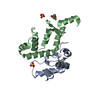
| |||||||||||||||
|---|---|---|---|---|---|---|---|---|---|---|---|---|---|---|---|---|
| 1 | 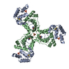
| |||||||||||||||
| Unit cell |
| |||||||||||||||
| Components on special symmetry positions |
|
-Components
-Protein , 2 types, 2 molecules AB
| #1: Protein | Mass: 10268.640 Da / Num. of mol.: 1 Source method: isolated from a genetically manipulated source Details: The first five residues are not observed in the final structure Source: (gene. exp.) Mycobacterium tuberculosis (bacteria) / Strain: ATCC 25618 / H37Rv / Gene: whiB3, Rv3416 / Production host: |
|---|---|
| #2: Protein | Mass: 12650.234 Da / Num. of mol.: 1 Source method: isolated from a genetically manipulated source Details: The 82 residues in the C-terminal of RNA polymerase sigma factor was fused with the flap tip helix (T815-A829) from RNA polymerase beta subunit, with six histidine tag at the N-terminus Source: (gene. exp.) Mycobacterium tuberculosis (bacteria) / Strain: ATCC 25618 / H37RvGene: sigA, mysA, rpoD, rpoV, Rv2703, MTCY05A6.24, rpoB, Rv0667, MTCI376.08c Production host: References: UniProt: P9WGI1, UniProt: P9WGY9, DNA-directed RNA polymerase |
-Non-polymers , 5 types, 112 molecules 


| #3: Chemical | ChemComp-SF4 /  Mass: 351.640 Da / Num. of mol.: 1 / Source method: obtained synthetically / Formula: Fe4S4 / Feature type: SUBJECT OF INVESTIGATION Mass: 351.640 Da / Num. of mol.: 1 / Source method: obtained synthetically / Formula: Fe4S4 / Feature type: SUBJECT OF INVESTIGATION | ||||||
|---|---|---|---|---|---|---|---|
| #4: Chemical |  Mass: 96.063 Da / Num. of mol.: 2 / Source method: obtained synthetically / Formula: SO4 Mass: 96.063 Da / Num. of mol.: 2 / Source method: obtained synthetically / Formula: SO4#5: Chemical | ChemComp-TRS / |  Mass: 122.143 Da / Num. of mol.: 1 / Source method: obtained synthetically / Formula: C4H12NO3 / Comment: pH buffer*YM Mass: 122.143 Da / Num. of mol.: 1 / Source method: obtained synthetically / Formula: C4H12NO3 / Comment: pH buffer*YM#6: Chemical |  Mass: 58.693 Da / Num. of mol.: 3 / Source method: obtained synthetically / Formula: Ni / Feature type: SUBJECT OF INVESTIGATION Mass: 58.693 Da / Num. of mol.: 3 / Source method: obtained synthetically / Formula: Ni / Feature type: SUBJECT OF INVESTIGATION#7: Water | ChemComp-HOH / | Mass: 18.015 Da / Num. of mol.: 105 / Source method: isolated from a natural source / Formula: H2O |
-Details
| Has ligand of interest | Y |
|---|
-Experimental details
-Experiment
| Experiment | Method: X-RAY DIFFRACTION / Number of used crystals: 1 |
|---|
- Sample preparation
| Crystal | Density Matthews: 1.96 Å3/Da / Density % sol: 37.4 % / Description: Tiny triangle-shaped crystals |
|---|---|
| Crystal grow | Temperature: 293 K / Method: vapor diffusion / pH: 8 / Details: 10-20 mM NiCl2, 0.8-1.0 M Li2SO4 / Temp details: Room temperature |
-Data collection
| Diffraction | Mean temperature: 100 K / Ambient temp details: Collect under Liquid Nitrogen stream / Serial crystal experiment: N |
|---|---|
| Diffraction source | Source: SYNCHROTRON / Site: SSRL / Beamline: BL12-2 / Wavelength: 0.9795 Å |
| Detector | Type: DECTRIS PILATUS 6M / Detector: PIXEL / Date: Feb 11, 2021 |
| Radiation | Monochromator: M / Protocol: SINGLE WAVELENGTH / Monochromatic (M) / Laue (L): M / Scattering type: x-ray |
| Radiation wavelength | Wavelength: 0.9795 Å / Relative weight: 1 |
| Reflection | Resolution: 1.5→50 Å / Num. obs: 27421 / % possible obs: 99.8 % / Redundancy: 18.1 % / Biso Wilson estimate: 26.01 Å2 / CC1/2: 1 / CC star: 1 / Rmerge(I) obs: 0.077 / Rpim(I) all: 0.018 / Rrim(I) all: 0.079 / Χ2: 1.092 / Net I/σ(I): 45.4 |
| Reflection shell | Resolution: 1.5→1.53 Å / Redundancy: 14.2 % / Rmerge(I) obs: 2.16 / Mean I/σ(I) obs: 1.6 / Num. unique obs: 2696 / CC1/2: 0.713 / CC star: 0.922 / Rpim(I) all: 0.578 / Rrim(I) all: 2.239 / Χ2: 0.575 / % possible all: 99.7 |
- Processing
| Software |
| |||||||||||||||||||||||||||||||||||||||||||||||||||||||||||||||||||||||||||
|---|---|---|---|---|---|---|---|---|---|---|---|---|---|---|---|---|---|---|---|---|---|---|---|---|---|---|---|---|---|---|---|---|---|---|---|---|---|---|---|---|---|---|---|---|---|---|---|---|---|---|---|---|---|---|---|---|---|---|---|---|---|---|---|---|---|---|---|---|---|---|---|---|---|---|---|---|
| Refinement | Method to determine structure: MOLECULAR REPLACEMENT Starting model: 8CWT Resolution: 1.5→34.87 Å / SU ML: 0.2003 / Cross valid method: FREE R-VALUE / σ(F): 1.96 / Phase error: 26.139 Stereochemistry target values: GeoStd + Monomer Library + CDL v1.2 Details: Refined with Phenix.refine GUI. Both B-factors and individual B-factors are refined.
| |||||||||||||||||||||||||||||||||||||||||||||||||||||||||||||||||||||||||||
| Solvent computation | Shrinkage radii: 0.9 Å / VDW probe radii: 1.11 Å / Solvent model: FLAT BULK SOLVENT MODEL | |||||||||||||||||||||||||||||||||||||||||||||||||||||||||||||||||||||||||||
| Displacement parameters | Biso mean: 41.31 Å2 | |||||||||||||||||||||||||||||||||||||||||||||||||||||||||||||||||||||||||||
| Refinement step | Cycle: LAST / Resolution: 1.5→34.87 Å
| |||||||||||||||||||||||||||||||||||||||||||||||||||||||||||||||||||||||||||
| Refine LS restraints |
| |||||||||||||||||||||||||||||||||||||||||||||||||||||||||||||||||||||||||||
| LS refinement shell |
| |||||||||||||||||||||||||||||||||||||||||||||||||||||||||||||||||||||||||||
| Refinement TLS params. | Method: refined / Refine-ID: X-RAY DIFFRACTION
| |||||||||||||||||||||||||||||||||||||||||||||||||||||||||||||||||||||||||||
| Refinement TLS group |
|
