 Movie
Movie Controller

[English] 日本語
 Yorodumi
- PDB-8ct7: Catalytic Core Domain of HIV-1 Integrase (F185K) bound with BI-224436 -
+ Open data

- Basic information
| Entry | Database: PDB / ID: 8ct7 | ||||||
|---|---|---|---|---|---|---|---|
| Title | Catalytic Core Domain of HIV-1 Integrase (F185K) bound with BI-224436 | ||||||
 Components Components | Integrase | ||||||
 Keywords Keywords | VIRAL PROTEIN/INHIBITOR / VIRAL DNA INTEGRATION / DNA BINDING / LEDGF BINDING / VIRAL PROTEIN / VIRAL PROTEIN-INHIBITOR complex | ||||||
| Function / homology |  Function and homology information Function and homology informationexoribonuclease H activity / DNA integration / viral genome integration into host DNA / establishment of integrated proviral latency / RNA stem-loop binding / viral penetration into host nucleus / host multivesicular body / RNA-directed DNA polymerase activity / RNA-DNA hybrid ribonuclease activity / host cell ...exoribonuclease H activity / DNA integration / viral genome integration into host DNA / establishment of integrated proviral latency / RNA stem-loop binding / viral penetration into host nucleus / host multivesicular body / RNA-directed DNA polymerase activity / RNA-DNA hybrid ribonuclease activity / host cell / viral nucleocapsid / DNA recombination / aspartic-type endopeptidase activity / DNA-directed DNA polymerase activity / symbiont-mediated suppression of host gene expression / viral translational frameshifting / symbiont entry into host cell / lipid binding / host cell nucleus / host cell plasma membrane / virion membrane / proteolysis / DNA binding / zinc ion binding Similarity search - Function | ||||||
| Biological species |   Human immunodeficiency virus 1 Human immunodeficiency virus 1 | ||||||
| Method |  X-RAY DIFFRACTION / SYNCHROTRON / MOLECULAR REPLACEMENT / Resolution: 2.13 Å X-RAY DIFFRACTION / SYNCHROTRON / MOLECULAR REPLACEMENT / Resolution: 2.13 Å | ||||||
 Authors Authors | Gupta, K. / Van Duyne, G.D. / Eilers, G. / Bushman, F.D. | ||||||
| Funding support |  United States, 1items United States, 1items
| ||||||
 Citation Citation | Journal: Plos Pathog. / Year: 2023 Title: Structure of a HIV-1 IN-Allosteric inhibitor complex at 2.93 angstrom resolution: Routes to inhibitor optimization. Authors: Eilers, G. / Gupta, K. / Allen, A. / Montermoso, S. / Murali, H. / Sharp, R. / Hwang, Y. / Bushman, F.D. / Van Duyne, G. | ||||||
| History |
|
- Structure visualization
| Structure viewer | Molecule: MolmilJmol/JSmol |
|---|
- Downloads & links
-Download
| PDBx/mmCIF format | 8ct7.cif.gz | 46.9 KB | Display | PDBx/mmCIF format |
|---|---|---|---|---|
| PDB format | pdb8ct7.ent.gz | 30.2 KB | Display | PDB format |
| PDBx/mmJSON format | 8ct7.json.gz | Tree view | PDBx/mmJSON format | |
| Others |  Other downloads Other downloads |
-Validation report
| Arichive directory | https://data.pdbj.org/pub/pdb/validation_reports/ct/8ct7ftp://data.pdbj.org/pub/pdb/validation_reports/ct/8ct7 | HTTPS FTP |
|---|
-Related structure data
| Related structure data |  8ct5C  8ctaC 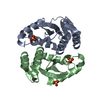 3l3uS S: Starting model for refinement C: citing same article ( |
|---|---|
| Similar structure data |
-Links
 PDBj
PDBj- Assembly
| Deposited unit | 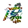
| ||||||||||
|---|---|---|---|---|---|---|---|---|---|---|---|
| 1 | 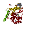
| ||||||||||
| Unit cell |
|
-Components
| #1: Protein | Mass: 17894.152 Da / Num. of mol.: 1 / Mutation: F185K Source method: isolated from a genetically manipulated source Source: (gene. exp.) Human immunodeficiency virus 1 / Gene: pol / Variant: F185K / Plasmid: pET24 / Production host:  |
|---|---|
| #2: Chemical | ChemComp-L3D / (  Mass: 442.506 Da / Num. of mol.: 1 / Source method: obtained synthetically / Formula: C27H26N2O4 / Feature type: SUBJECT OF INVESTIGATION Mass: 442.506 Da / Num. of mol.: 1 / Source method: obtained synthetically / Formula: C27H26N2O4 / Feature type: SUBJECT OF INVESTIGATION |
| #3: Chemical | ChemComp-EDO /   Mass: 62.068 Da / Num. of mol.: 1 / Source method: obtained synthetically / Formula: C2H6O2 Mass: 62.068 Da / Num. of mol.: 1 / Source method: obtained synthetically / Formula: C2H6O2 |
| #4: Chemical | ChemComp-SO4 /   Mass: 96.063 Da / Num. of mol.: 1 / Source method: obtained synthetically / Formula: SO4 Mass: 96.063 Da / Num. of mol.: 1 / Source method: obtained synthetically / Formula: SO4 |
| #5: Water | ChemComp-HOH /  Mass: 18.015 Da / Num. of mol.: 19 / Source method: isolated from a natural source / Formula: H2O Mass: 18.015 Da / Num. of mol.: 19 / Source method: isolated from a natural source / Formula: H2O |
| Has ligand of interest | Y |
| Has protein modification | Y |
-Experimental details
-Experiment
| Experiment | Method: X-RAY DIFFRACTION / Number of used crystals: 1 |
|---|
- Sample preparation
| Crystal | Density Matthews: 2.82 Å3/Da / Density % sol: 56.33 % |
|---|---|
| Crystal grow | Temperature: 293.15 K / Method: vapor diffusion, hanging drop / pH: 7.5 Details: 7% PEG 8000, 0.2 M ammonium sulfate, 0.1 M sodium cacodylate, pH 6.5, 5 mM manganese chloride, 5 mM magnesium chloride, and 5 mM DTT |
-Data collection
| Diffraction | Mean temperature: 100 K / Serial crystal experiment: N |
|---|---|
| Diffraction source | Source: SYNCHROTRON / Site: CHESS / Beamline: A1 / Wavelength: 0.92 Å |
| Detector | Type: DECTRIS EIGER2 X 16M / Detector: PIXEL / Date: Oct 13, 2019 |
| Radiation | Protocol: SINGLE WAVELENGTH / Monochromatic (M) / Laue (L): M / Scattering type: x-ray |
| Radiation wavelength | Wavelength: 0.92 Å / Relative weight: 1 |
| Reflection | Resolution: 2.13→29.38 Å / Num. obs: 11625 / % possible obs: 99.9 % / Redundancy: 2 % / Biso Wilson estimate: 44.8 Å2 / CC1/2: 1 / CC star: 1 / Rmerge(I) obs: 0.017 / Rpim(I) all: 0.017 / Rrim(I) all: 0.024 / Net I/σ(I): 29.5 |
| Reflection shell | Resolution: 2.13→2.206 Å / Redundancy: 2 % / Rmerge(I) obs: 0.16 / Num. unique obs: 1133 / CC1/2: 0.927 / CC star: 0.981 / Rpim(I) all: 0.017 / Rrim(I) all: 0.024 / % possible all: 100 |
- Processing
| Software |
| ||||||||||||||||||||
|---|---|---|---|---|---|---|---|---|---|---|---|---|---|---|---|---|---|---|---|---|---|
| Refinement | Method to determine structure: MOLECULAR REPLACEMENT Starting model: 3L3U Resolution: 2.13→29.38 Å / SU B: 4.38 / SU ML: 0.114 / Cross valid method: THROUGHOUT / ESU R: 0.2 / ESU R Free: 0.168 / Details: HYDROGENS HAVE BEEN ADDED IN THE RIDING POSITIONS
| ||||||||||||||||||||
| Displacement parameters | Biso mean: 50.584 Å2
| ||||||||||||||||||||
| Refinement step | Cycle: LAST / Resolution: 2.13→29.38 Å
|
