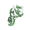
- PDB-8cri: Crystal structure of LplA1 in complex with lipoic acid (Listeria ... -
+
Open data
ID or keywords:
Loading...
-
Basic information
Entry
Database: PDB / ID: 8cri
Title
Crystal structure of LplA1 in complex with lipoic acid (Listeria monocytogenes)
Components
lipoate--protein ligase
Keywords
LIGASE / LIPOATE / SALVAGE / ANTI-INFECTIVES / DRUG DEVELOPMENT
Function / homology
Function and homology information
lipoyltransferase activity / lipoate-protein ligase / lipoate-protein ligase activity / protein lipoylation / ATP binding / metal ion binding / cytoplasm Similarity search - Function
Lipoate protein ligase, C-terminal / Bacterial lipoate protein ligase C-terminus / Lipoyltransferase/lipoate-protein ligase / Lipoyl protein ligase A/B catalytic domain / CO dehydrogenase flavoprotein, C-terminal domain / Biotinyl protein ligase (BPL) and lipoyl protein ligase (LPL) catalytic domain profile. / Biotinyl protein ligase (BPL) and lipoyl protein ligase (LPL), catalytic domain / Bira Bifunctional Protein; Domain 2 / BirA Bifunctional Protein; domain 2 / Enolase-like; domain 1 ...Lipoate protein ligase, C-terminal / Bacterial lipoate protein ligase C-terminus / Lipoyltransferase/lipoate-protein ligase / Lipoyl protein ligase A/B catalytic domain / CO dehydrogenase flavoprotein, C-terminal domain / Biotinyl protein ligase (BPL) and lipoyl protein ligase (LPL) catalytic domain profile. / Biotinyl protein ligase (BPL) and lipoyl protein ligase (LPL), catalytic domain / Bira Bifunctional Protein; Domain 2 / BirA Bifunctional Protein; domain 2 / Enolase-like; domain 1 / Class II Aminoacyl-tRNA synthetase/Biotinyl protein ligase (BPL) and lipoyl protein ligase (LPL) / 2-Layer Sandwich / Alpha Beta Similarity search - Domain/homology
Type: DECTRIS EIGER X 16M / Detector: PIXEL / Date: Oct 15, 2022
Radiation
Protocol: SINGLE WAVELENGTH / Monochromatic (M) / Laue (L): M / Scattering type: x-ray
Radiation wavelength
Wavelength: 1 Å / Relative weight: 1
Reflection
Resolution: 2.1→30 Å / Num. obs: 44270 / % possible obs: 97.1 % / Redundancy: 2.9 % / Rmerge(I) obs: 0.055 / Net I/σ(I): 10.7
Reflection shell
Resolution: 2.1→2.2 Å / Redundancy: 3 % / Rmerge(I) obs: 0.66 / Mean I/σ(I) obs: 2.1 / Num. unique obs: 5813 / % possible all: 98.9
-
Processing
Software
Name
Version
Classification
REFMAC
5.8.0267
refinement
XDS
datareduction
XSCALE
datascaling
PHASER
phasing
Refinement
Method to determine structure: MOLECULAR REPLACEMENT / Resolution: 2.1→30 Å / Cor.coef. Fo:Fc: 0.945 / Cor.coef. Fo:Fc free: 0.929 / SU B: 16.188 / SU ML: 0.18 / Cross valid method: THROUGHOUT / ESU R Free: 0.185 / Stereochemistry target values: MAXIMUM LIKELIHOOD / Details: HYDROGENS HAVE BEEN ADDED IN THE RIDING POSITIONS
Rfactor
Num. reflection
% reflection
Selection details
Rfree
0.25145
2213
5 %
RANDOM
Rwork
0.22334
-
-
-
obs
0.22487
42047
97.11 %
-
Solvent computation
Ion probe radii: 0.8 Å / Shrinkage radii: 0.8 Å / VDW probe radii: 1.2 Å / Solvent model: MASK
 Movie
Movie Controller

 Yorodumi
 Open data
 Basic information
 Components
 Keywords
 Function and homology information
 Listeria monocytogenes (bacteria)
 X-RAY DIFFRACTION /
  Authors
 Germany, 1items
  Citation
 Structure visualization
 Downloads & links
 Other downloads
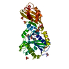

 PDBj
PDBj
 Assembly
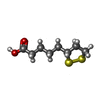


 Mass: 206.326 Da / Num. of mol.: 1 / Source method: obtained synthetically / Formula: C8H14O2S2 / Feature type: SUBJECT OF INVESTIGATION
 Mass: 35.453 Da / Num. of mol.: 1 / Source method: obtained synthetically / Formula: Cl
 Mass: 96.063 Da / Num. of mol.: 1 / Source method: obtained synthetically / Formula: SO4
 Mass: 62.068 Da / Num. of mol.: 1 / Source method: obtained synthetically / Formula: C2H6O2
 Sample preparation
 / Beamline: X06SA / Wavelength: 1 Å
 Processing