 Movie
Movie Controller


+ Open data

- Basic information
| Entry | Database: PDB / ID: 8cqd | ||||||
|---|---|---|---|---|---|---|---|
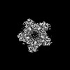
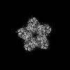
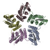
| Title | Cryo-EM structure of hexameric proteorhodopsin A18L mutant | ||||||
 Components Components | Green-light absorbing proteorhodopsin | ||||||
 Keywords Keywords | PROTON TRANSPORT / Membrane protein / Light-driven proton pump / Proteorhodopsin | ||||||
| Function / homology |  Function and homology information Function and homology informationlight-activated monoatomic ion channel activity / photoreceptor activity / phototransduction / plasma membrane Similarity search - Function | ||||||
| Biological species |  uncultured Gammaproteobacteria bacterium (environmental samples) uncultured Gammaproteobacteria bacterium (environmental samples) | ||||||
| Method | ELECTRON MICROSCOPY / single particle reconstruction / cryo EM / Resolution: 3.54 Å | ||||||
 Authors Authors | Hirschi, S. / Lemmin, T. / Fotiadis, D. | ||||||
| Funding support |  Switzerland, 1items Switzerland, 1items
| ||||||
 Citation Citation | Journal: Nat Commun / Year: 2024 Title: Structural insights into the mechanism and dynamics of proteorhodopsin biogenesis and retinal scavenging. Authors: Stephan Hirschi / Thomas Lemmin / Nooraldeen Ayoub / David Kalbermatter / Daniele Pellegata / Zöhre Ucurum / Jürg Gertsch / Dimitrios Fotiadis /  Abstract: Microbial ion-pumping rhodopsins (MRs) are extensively studied retinal-binding membrane proteins. However, their biogenesis, including oligomerisation and retinal incorporation, remains poorly ...Microbial ion-pumping rhodopsins (MRs) are extensively studied retinal-binding membrane proteins. However, their biogenesis, including oligomerisation and retinal incorporation, remains poorly understood. The bacterial green-light absorbing proton pump proteorhodopsin (GPR) has emerged as a model protein for MRs and is used here to address these open questions using cryo-electron microscopy (cryo-EM) and molecular dynamics (MD) simulations. Specifically, conflicting studies regarding GPR stoichiometry reported pentamer and hexamer mixtures without providing possible assembly mechanisms. We report the pentameric and hexameric cryo-EM structures of a GPR mutant, uncovering the role of the unprocessed N-terminal signal peptide in the assembly of hexameric GPR. Furthermore, certain proteorhodopsin-expressing bacteria lack retinal biosynthesis pathways, suggesting that they scavenge the cofactor from their environment. We shed light on this hypothesis by solving the cryo-EM structure of retinal-free proteoopsin, which together with mass spectrometry and MD simulations suggests that decanoate serves as a temporary placeholder for retinal in the chromophore binding pocket. Further MD simulations elucidate possible pathways for the exchange of decanoate and retinal, offering a mechanism for retinal scavenging. Collectively, our findings provide insights into the biogenesis of MRs, including their oligomeric assembly, variations in protomer stoichiometry and retinal incorporation through a potential cofactor scavenging mechanism. | ||||||
| History |
|
- Structure visualization
| Structure viewer | Molecule: MolmilJmol/JSmol |
|---|
- Downloads & links
-Download
| PDBx/mmCIF format | 8cqd.cif.gz | 239.5 KB | Display | PDBx/mmCIF format |
|---|---|---|---|---|
| PDB format | pdb8cqd.ent.gz | 195.1 KB | Display | PDB format |
| PDBx/mmJSON format | 8cqd.json.gz | Tree view | PDBx/mmJSON format | |
| Others |  Other downloads Other downloads |
-Validation report
| Summary document | 8cqd_validation.pdf.gz | 1.4 MB | Display | wwPDB validaton report |
|---|---|---|---|---|
| Full document | 8cqd_full_validation.pdf.gz | 1.4 MB | Display | |
| Data in XML | 8cqd_validation.xml.gz | 42 KB | Display | |
| Data in CIF | 8cqd_validation.cif.gz | 61 KB | Display | |
| Arichive directory | https://data.pdbj.org/pub/pdb/validation_reports/cq/8cqdftp://data.pdbj.org/pub/pdb/validation_reports/cq/8cqd | HTTPS FTP |
-Related structure data
| Related structure data | 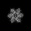 16796MC 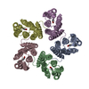 8cnkC 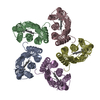 8cqcC C: citing same article ( M: map data used to model this data |
|---|---|
| Similar structure data |
-Links
 PDBj
PDBj


- Assembly
| Deposited unit | 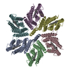
|
|---|---|
| 1 |
|
-Components
| #1: Protein | Mass: 28110.854 Da / Num. of mol.: 6 / Mutation: A18L Source method: isolated from a genetically manipulated source Source: (gene. exp.) uncultured Gammaproteobacteria bacterium (environmental samples)Production host: #2: Chemical | ChemComp-RET /   Mass: 284.436 Da / Num. of mol.: 6 / Source method: obtained synthetically / Formula: C20H28O / Feature type: SUBJECT OF INVESTIGATION Mass: 284.436 Da / Num. of mol.: 6 / Source method: obtained synthetically / Formula: C20H28O / Feature type: SUBJECT OF INVESTIGATIONHas ligand of interest | Y | Has protein modification | Y | |
|---|
-Experimental details
-Experiment
| Experiment | Method: ELECTRON MICROSCOPY |
|---|---|
| EM experiment | Aggregation state: PARTICLE / 3D reconstruction method: single particle reconstruction |
- Sample preparation
| Component | Name: Hexameric proteorhodopsin A18L / Type: COMPLEX / Entity ID: #1 / Source: RECOMBINANT |
|---|---|
| Molecular weight | Experimental value: NO |
| Source (natural) | Organism: uncultured Gammaproteobacteria bacterium (environmental samples) |
| Source (recombinant) | Organism: |
| Buffer solution | pH: 7.5 |
| Specimen | Conc.: 4.2 mg/ml / Embedding applied: NO / Shadowing applied: NO / Staining applied: NO / Vitrification applied: YES |
| Vitrification | Instrument: FEI VITROBOT MARK IV / Cryogen name: ETHANE |
- Electron microscopy imaging
| Experimental equipment |  Model: Titan Krios / Image courtesy: FEI Company |
|---|---|
| Microscopy | Model: FEI TITAN KRIOS |
| Electron gun | Electron source:  FIELD EMISSION GUN / Accelerating voltage: 300 kV / Illumination mode: OTHER FIELD EMISSION GUN / Accelerating voltage: 300 kV / Illumination mode: OTHER |
| Electron lens | Mode: BRIGHT FIELD / Nominal magnification: 105000 X / Nominal defocus max: 1700 nm / Nominal defocus min: 700 nm |
| Image recording | Electron dose: 49.8 e/Å2 / Film or detector model: GATAN K3 (6k x 4k) |
- Processing
| EM software | Name: cryoSPARC / Category: 3D reconstruction | ||||||||||||||||||||||||
|---|---|---|---|---|---|---|---|---|---|---|---|---|---|---|---|---|---|---|---|---|---|---|---|---|---|
| CTF correction | Type: PHASE FLIPPING AND AMPLITUDE CORRECTION | ||||||||||||||||||||||||
| Symmetry | Point symmetry: C2 (2 fold cyclic) | ||||||||||||||||||||||||
| 3D reconstruction | Resolution: 3.54 Å / Resolution method: FSC 0.143 CUT-OFF / Num. of particles: 237013 / Symmetry type: POINT | ||||||||||||||||||||||||
| Refine LS restraints |
|
