 Movie
Movie Controller

[English] 日本語
 Yorodumi
- EMDB-16759: Cryo-EM structure of retinal-free proteoopsin bound to decanoate -
+ Open data

- Basic information
| Entry | 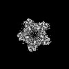 | |||||||||
|---|---|---|---|---|---|---|---|---|---|---|
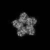
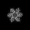
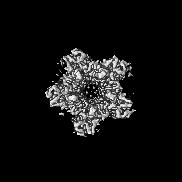
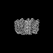
| Title | Cryo-EM structure of retinal-free proteoopsin bound to decanoate | |||||||||
 Map data Map data | ||||||||||
 Sample Sample |
| |||||||||
 Keywords Keywords | Membrane protein / Light-driven proton pump / Proteorhodopsin / Proteoopsin / PROTON TRANSPORT | |||||||||
| Function / homology |  Function and homology information Function and homology informationlight-activated monoatomic ion channel activity / phototransduction / photoreceptor activity / plasma membrane Similarity search - Function | |||||||||
| Biological species |  uncultured Gammaproteobacteria bacterium (environmental samples) uncultured Gammaproteobacteria bacterium (environmental samples) | |||||||||
| Method | single particle reconstruction / cryo EM / Resolution: 2.97 Å | |||||||||
 Authors Authors | Hirschi S / Lemmin T / Fotiadis D | |||||||||
| Funding support |  Switzerland, 1 items Switzerland, 1 items
| |||||||||
 Citation Citation | Journal: Nat Commun / Year: 2024 Title: Structural insights into the mechanism and dynamics of proteorhodopsin biogenesis and retinal scavenging. Authors: Stephan Hirschi / Thomas Lemmin / Nooraldeen Ayoub / David Kalbermatter / Daniele Pellegata / Zöhre Ucurum / Jürg Gertsch / Dimitrios Fotiadis /  Abstract: Microbial ion-pumping rhodopsins (MRs) are extensively studied retinal-binding membrane proteins. However, their biogenesis, including oligomerisation and retinal incorporation, remains poorly ...Microbial ion-pumping rhodopsins (MRs) are extensively studied retinal-binding membrane proteins. However, their biogenesis, including oligomerisation and retinal incorporation, remains poorly understood. The bacterial green-light absorbing proton pump proteorhodopsin (GPR) has emerged as a model protein for MRs and is used here to address these open questions using cryo-electron microscopy (cryo-EM) and molecular dynamics (MD) simulations. Specifically, conflicting studies regarding GPR stoichiometry reported pentamer and hexamer mixtures without providing possible assembly mechanisms. We report the pentameric and hexameric cryo-EM structures of a GPR mutant, uncovering the role of the unprocessed N-terminal signal peptide in the assembly of hexameric GPR. Furthermore, certain proteorhodopsin-expressing bacteria lack retinal biosynthesis pathways, suggesting that they scavenge the cofactor from their environment. We shed light on this hypothesis by solving the cryo-EM structure of retinal-free proteoopsin, which together with mass spectrometry and MD simulations suggests that decanoate serves as a temporary placeholder for retinal in the chromophore binding pocket. Further MD simulations elucidate possible pathways for the exchange of decanoate and retinal, offering a mechanism for retinal scavenging. Collectively, our findings provide insights into the biogenesis of MRs, including their oligomeric assembly, variations in protomer stoichiometry and retinal incorporation through a potential cofactor scavenging mechanism. | |||||||||
| History |
|
- Structure visualization
| Supplemental images |
|---|

- Downloads & links
-EMDB archive
| Map data | emd_16759.map.gz | 20.3 MB | EMDB map data format | |
|---|---|---|---|---|
| Header (meta data) | emd-16759-v30.xmlemd-16759.xml | 14.2 KB 14.2 KB | Display Display | EMDB header |
| Images |  emd_16759.png emd_16759.png | 166.1 KB | ||
| Filedesc metadata | emd-16759.cif.gz | 5.3 KB | ||
| Others | emd_16759_half_map_1.map.gzemd_16759_half_map_2.map.gz | 20.9 MB 20.9 MB | ||
| Archive directory |  http://ftp.pdbj.org/pub/emdb/structures/EMD-16759ftp://ftp.pdbj.org/pub/emdb/structures/EMD-16759 http://ftp.pdbj.org/pub/emdb/structures/EMD-16759ftp://ftp.pdbj.org/pub/emdb/structures/EMD-16759 | HTTPS FTP |
-Related structure data
| Related structure data |  8cnkMC  8cqcC  8cqdC M: atomic model generated by this map C: citing same article ( |
|---|---|
| Similar structure data |
-Links
| EMDB pages | EMDB (EBI/PDBe) / EMDataResource |
|---|---|
| Related items in Molecule of the Month |


-Map
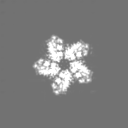
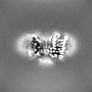
| File | Download / File: emd_16759.map.gz / Format: CCP4 / Size: 23 MB / Type: IMAGE STORED AS FLOATING POINT NUMBER (4 BYTES) | ||||||||||||||||||||||||||||||||||||
|---|---|---|---|---|---|---|---|---|---|---|---|---|---|---|---|---|---|---|---|---|---|---|---|---|---|---|---|---|---|---|---|---|---|---|---|---|---|
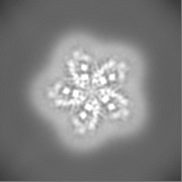
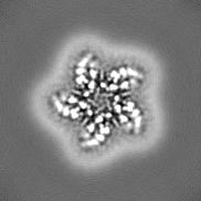
| Projections & slices | Image control
Images are generated by Spider. | ||||||||||||||||||||||||||||||||||||
| Voxel size | X=Y=Z: 0.99231 Å | ||||||||||||||||||||||||||||||||||||
| Density |
| ||||||||||||||||||||||||||||||||||||
| Symmetry | Space group: 1 | ||||||||||||||||||||||||||||||||||||
| Details | EMDB XML:
|
 Z (Sec.)
 Y (Row.)
 X (Col.)


-Supplemental data
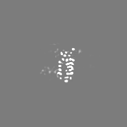
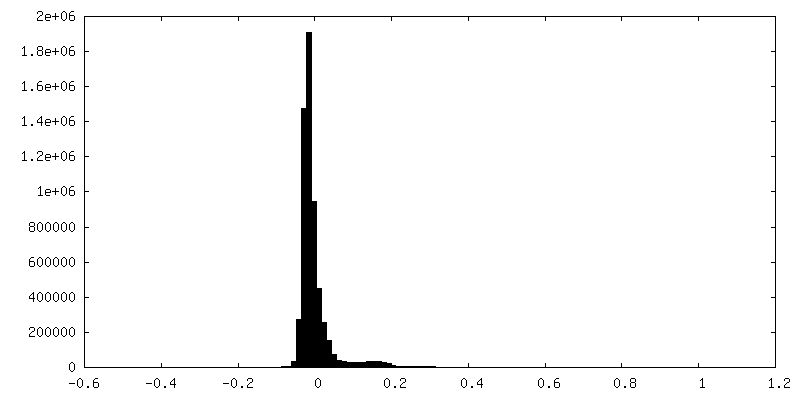
-Half map: #2
| File | emd_16759_half_map_1.map | ||||||||||||
|---|---|---|---|---|---|---|---|---|---|---|---|---|---|
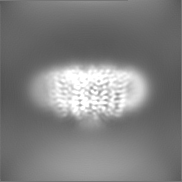
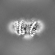
| Projections & Slices |
| ||||||||||||
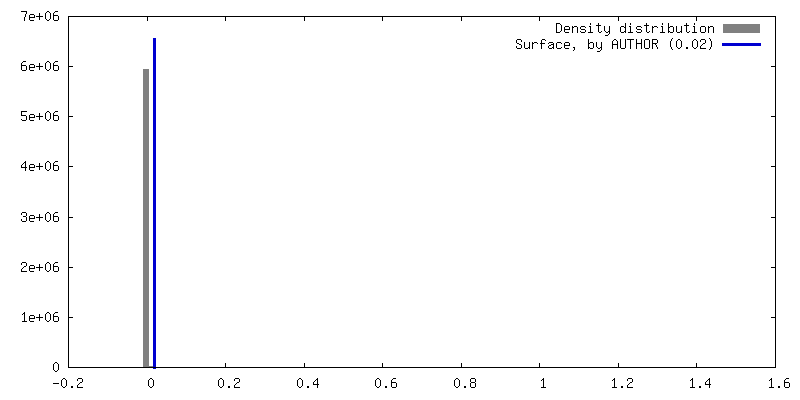
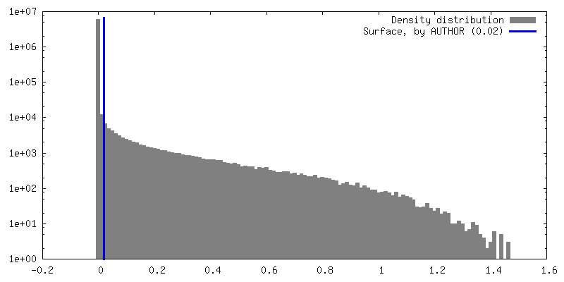
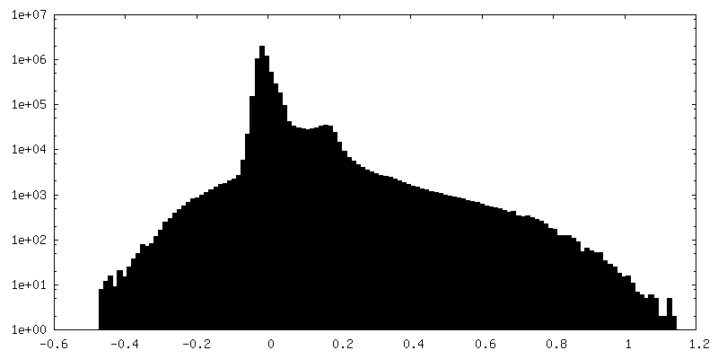
| Density Histograms |


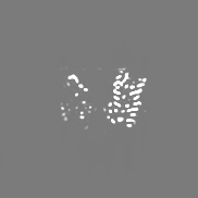
-Half map: #1
| File | emd_16759_half_map_2.map | ||||||||||||
|---|---|---|---|---|---|---|---|---|---|---|---|---|---|
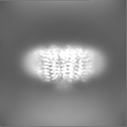
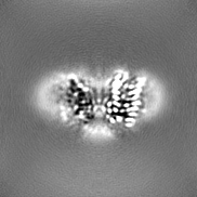
| Projections & Slices |
| ||||||||||||
| Density Histograms |


- Sample components
-Entire : Pentameric proteoopsin bound to decanoate
| Entire | Name: Pentameric proteoopsin bound to decanoate |
|---|---|
| Components |
|
-Supramolecule #1: Pentameric proteoopsin bound to decanoate
| Supramolecule | Name: Pentameric proteoopsin bound to decanoate / type: complex / ID: 1 / Parent: 0 / Macromolecule list: #1 |
|---|---|
| Source (natural) | Organism: uncultured Gammaproteobacteria bacterium (environmental samples) |
-Macromolecule #1: Green-light absorbing proteorhodopsin
| Macromolecule | Name: Green-light absorbing proteorhodopsin / type: protein_or_peptide / ID: 1 / Number of copies: 5 / Enantiomer: LEVO |
|---|---|
| Source (natural) | Organism: uncultured Gammaproteobacteria bacterium (environmental samples) |
| Molecular weight | Theoretical: 26.359627 KDa |
| Recombinant expression | Organism: |
| Sequence | String: MAGGGDLDAS DYTGVSFWLV TAALLASTVF FFVERDRVSA KWKTSLTVSG LVTGIAFWHY MYMRGVWIET GDSPTVFRYI DWLLTVPLL ICEFYLILAA ATNVAGSLFK KLLVGSLVML VFGYMGEAGI MAAWPAFIIG CLAWVYMIYE LWAGEGKSAC N TASPAVQS ...String: MAGGGDLDAS DYTGVSFWLV TAALLASTVF FFVERDRVSA KWKTSLTVSG LVTGIAFWHY MYMRGVWIET GDSPTVFRYI DWLLTVPLL ICEFYLILAA ATNVAGSLFK KLLVGSLVML VFGYMGEAGI MAAWPAFIIG CLAWVYMIYE LWAGEGKSAC N TASPAVQS AYNTMMYIII FGWAIYPVGY FTGYLMGDGG SALNLNLIYN LADFVNKILF GLIIWNVAVK ESSNAGHHHH H UniProtKB: Green-light absorbing proteorhodopsin |
-Macromolecule #2: DECANOIC ACID
| Macromolecule | Name: DECANOIC ACID / type: ligand / ID: 2 / Number of copies: 5 / Formula: DKA |
|---|---|
| Molecular weight | Theoretical: 172.265 Da |
| Chemical component information |  ChemComp-DKA: |
-Experimental details
-Structure determination
| Method | cryo EM |
|---|---|
 Processing Processing | single particle reconstruction |
| Aggregation state | particle |
-Sample preparation
| Concentration | 4.0 mg/mL |
|---|---|
| Buffer | pH: 7.5 |
| Vitrification | Cryogen name: ETHANE / Instrument: FEI VITROBOT MARK IV |
- Electron microscopy
| Microscope | FEI TITAN KRIOS |
|---|---|
| Image recording | Film or detector model: GATAN K3 (6k x 4k) / Average electron dose: 49.8 e/Å2 |
| Electron beam | Acceleration voltage: 300 kV / Electron source:  FIELD EMISSION GUN FIELD EMISSION GUN |
| Electron optics | Illumination mode: OTHER / Imaging mode: BRIGHT FIELD / Nominal defocus max: 1.7 µm / Nominal defocus min: 0.7000000000000001 µm / Nominal magnification: 130000 |
| Experimental equipment |  Model: Titan Krios / Image courtesy: FEI Company |
-Image processing
| Startup model | Type of model: NONE |
|---|---|
| Final reconstruction | Applied symmetry - Point group: C5 (5 fold cyclic) / Algorithm: BACK PROJECTION / Resolution.type: BY AUTHOR / Resolution: 2.97 Å / Resolution method: FSC 0.143 CUT-OFF / Software - Name: cryoSPARC / Number images used: 947286 |
| Initial angle assignment | Type: ANGULAR RECONSTITUTION |
| Final angle assignment | Type: ANGULAR RECONSTITUTION |
