 Movie
Movie Controller

[English] 日本語
 Yorodumi
- PDB-8cq4: Bifunctional cyclohexadienyl dehydratase/chorismate mutase from J... -
+ Open data

- Basic information
| Entry | Database: PDB / ID: 8cq4 | ||||||
|---|---|---|---|---|---|---|---|
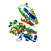
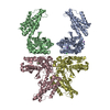
| Title | Bifunctional cyclohexadienyl dehydratase/chorismate mutase from Janthinobacterium sp. HH01 | ||||||
 Components Components | Bifunctional cyclohexadienyl dehydratase/chorismate mutase from Janthinobacterium sp. HH01 | ||||||
 Keywords Keywords | UNKNOWN FUNCTION / chorismate mutase / cyclohexadienyl dehydratase / chorismate mutase/cyclohexadienyl dehydratase / cyclohexadienyl dehydratase/chorismate mutase / bifunctional chorismate mutase / bifunctional cyclohexadienyl dehydratase / bifunctional enzyme / shikimate pathway enzymes / metabolic enzymes / aromatic amino acid synthesis / protein crystal structure | ||||||
| Biological species |  Janthinobacterium sp. HH01 (bacteria) Janthinobacterium sp. HH01 (bacteria) | ||||||
| Method |  X-RAY DIFFRACTION / SYNCHROTRON / MOLECULAR REPLACEMENT / Resolution: 1.65 Å X-RAY DIFFRACTION / SYNCHROTRON / MOLECULAR REPLACEMENT / Resolution: 1.65 Å | ||||||
 Authors Authors | Khatanbaatar, T. / Cordara, G. / Krengel, U. | ||||||
| Funding support |  Switzerland, 1items Switzerland, 1items
| ||||||
 Citation Citation | Journal: J.Biol.Chem. / Year: 2023 Title: Novel exported fusion enzymes with chorismate mutase and cyclohexadienyl dehydratase activity: Shikimate pathway enzymes teamed up in no man's land. Authors: Stocker, C. / Khatanbaatar, T. / Bressan, L. / Wurth-Roderer, K. / Cordara, G. / Krengel, U. / Kast, P. | ||||||
| History |
|
- Structure visualization
| Structure viewer | Molecule:  MolmilJmol/JSmol MolmilJmol/JSmol |
|---|
- Downloads & links
-Download
| PDBx/mmCIF format | 8cq4.cif.gz | 105.7 KB | Display | PDBx/mmCIF format |
|---|---|---|---|---|
| PDB format | pdb8cq4.ent.gz | 77.5 KB | Display | PDB format |
| PDBx/mmJSON format | 8cq4.json.gz | Tree view | PDBx/mmJSON format | |
| Others |  Other downloads Other downloads |
-Validation report
| Summary document | 8cq4_validation.pdf.gz | 781.7 KB | Display | wwPDB validaton report |
|---|---|---|---|---|
| Full document | 8cq4_full_validation.pdf.gz | 786.7 KB | Display | |
| Data in XML | 8cq4_validation.xml.gz | 17.8 KB | Display | |
| Data in CIF | 8cq4_validation.cif.gz | 26.1 KB | Display | |
| Arichive directory | https://data.pdbj.org/pub/pdb/validation_reports/cq/8cq4ftp://data.pdbj.org/pub/pdb/validation_reports/cq/8cq4 | HTTPS FTP |
-Related structure data
-Links
 PDBj
PDBj- Assembly
| Deposited unit | 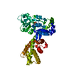
| ||||||||
|---|---|---|---|---|---|---|---|---|---|
| 1 |
| ||||||||
| Unit cell |
|
-Components
| #1: Protein | Mass: 47051.719 Da / Num. of mol.: 1 Source method: isolated from a genetically manipulated source Details: MES: 2-(N-morpholino)ethanesulfonic acid / Source: (gene. exp.) Janthinobacterium sp. HH01 (bacteria) / Gene: ELX09769.1 / Production host: |
|---|---|
| #2: Chemical | ChemComp-MES /   Mass: 195.237 Da / Num. of mol.: 1 / Source method: obtained synthetically / Formula: C6H13NO4S / Feature type: SUBJECT OF INVESTIGATION / Comment: pH buffer*YM Mass: 195.237 Da / Num. of mol.: 1 / Source method: obtained synthetically / Formula: C6H13NO4S / Feature type: SUBJECT OF INVESTIGATION / Comment: pH buffer*YM |
| #3: Water | ChemComp-HOH /  Mass: 18.015 Da / Num. of mol.: 154 / Source method: isolated from a natural source / Formula: H2O Mass: 18.015 Da / Num. of mol.: 154 / Source method: isolated from a natural source / Formula: H2O |
| Has ligand of interest | Y |
| Has protein modification | Y |
-Experimental details
-Experiment
| Experiment | Method: X-RAY DIFFRACTION / Number of used crystals: 1 |
|---|
- Sample preparation
| Crystal | Density Matthews: 2.19 Å3/Da / Density % sol: 43.8 % |
|---|---|
| Crystal grow | Temperature: 293.15 K / Method: vapor diffusion, sitting drop / pH: 6.5 Details: 10% w/v PEG 20 000 20% v/v PEG MME 550 0.03 M Sodium nitrate 0.03 M Disodium hydrogen phosphate 0.03 M Ammonium sulfate 0.1 M MES/imidazole pH 6.5 (Morpheus buffer 1) 3.5 mg/mL protein in 20 mM TRIS-HCl, pH 8 |
-Data collection
| Diffraction | Mean temperature: 100 K / Serial crystal experiment: N |
|---|---|
| Diffraction source | Source: SYNCHROTRON / Site: MAX IV  / Beamline: BioMAX / Wavelength: 0.976 Å / Beamline: BioMAX / Wavelength: 0.976 Å |
| Detector | Type: DECTRIS EIGER X 16M / Detector: PIXEL / Date: Dec 5, 2019 |
| Radiation | Protocol: SINGLE WAVELENGTH / Monochromatic (M) / Laue (L): M / Scattering type: x-ray |
| Radiation wavelength | Wavelength: 0.976 Å / Relative weight: 1 |
| Reflection | Resolution: 1.65→65.6 Å / Num. obs: 35076 / % possible obs: 93 % / Redundancy: 12.7 % / CC1/2: 0.998 / Net I/σ(I): 14.1 |
| Reflection shell | Resolution: 1.65→1.83 Å / Redundancy: 9.9 % / Mean I/σ(I) obs: 1.6 / Num. unique obs: 1754 / CC1/2: 0.542 / % possible all: 54.6 |
- Processing
| Software |
| |||||||||||||||||||||||||||||||||||||||||||||||||||||||||||||||||||||||||||||||||||||||||||||||||||||||||||||||||||||||||||||||||||||||||||||||||||||||||||
|---|---|---|---|---|---|---|---|---|---|---|---|---|---|---|---|---|---|---|---|---|---|---|---|---|---|---|---|---|---|---|---|---|---|---|---|---|---|---|---|---|---|---|---|---|---|---|---|---|---|---|---|---|---|---|---|---|---|---|---|---|---|---|---|---|---|---|---|---|---|---|---|---|---|---|---|---|---|---|---|---|---|---|---|---|---|---|---|---|---|---|---|---|---|---|---|---|---|---|---|---|---|---|---|---|---|---|---|---|---|---|---|---|---|---|---|---|---|---|---|---|---|---|---|---|---|---|---|---|---|---|---|---|---|---|---|---|---|---|---|---|---|---|---|---|---|---|---|---|---|---|---|---|---|---|---|---|
| Refinement | Method to determine structure: MOLECULAR REPLACEMENT / Resolution: 1.65→65.6 Å / Cor.coef. Fo:Fc: 0.959 / Cor.coef. Fo:Fc free: 0.93 / SU B: 3.091 / SU ML: 0.101 / Cross valid method: FREE R-VALUE / ESU R: 0.156 / ESU R Free: 0.15 Details: Hydrogens have been added in their riding positions
| |||||||||||||||||||||||||||||||||||||||||||||||||||||||||||||||||||||||||||||||||||||||||||||||||||||||||||||||||||||||||||||||||||||||||||||||||||||||||||
| Solvent computation | Ion probe radii: 0.8 Å / Shrinkage radii: 0.8 Å / VDW probe radii: 1.2 Å / Solvent model: MASK BULK SOLVENT | |||||||||||||||||||||||||||||||||||||||||||||||||||||||||||||||||||||||||||||||||||||||||||||||||||||||||||||||||||||||||||||||||||||||||||||||||||||||||||
| Displacement parameters | Biso mean: 28.766 Å2
| |||||||||||||||||||||||||||||||||||||||||||||||||||||||||||||||||||||||||||||||||||||||||||||||||||||||||||||||||||||||||||||||||||||||||||||||||||||||||||
| Refinement step | Cycle: LAST / Resolution: 1.65→65.6 Å
| |||||||||||||||||||||||||||||||||||||||||||||||||||||||||||||||||||||||||||||||||||||||||||||||||||||||||||||||||||||||||||||||||||||||||||||||||||||||||||
| Refine LS restraints |
| |||||||||||||||||||||||||||||||||||||||||||||||||||||||||||||||||||||||||||||||||||||||||||||||||||||||||||||||||||||||||||||||||||||||||||||||||||||||||||
| LS refinement shell |
|
