 Movie
Movie Controller

[English] 日本語
 Yorodumi
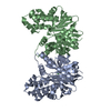
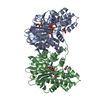
- PDB-8cp9: 1,6-anhydro-n-actetylmuramic acid kinase (AnmK)in complex with no... -
+ Open data

- Basic information
| Entry | Database: PDB / ID: 8cp9 | ||||||
|---|---|---|---|---|---|---|---|
| Title | 1,6-anhydro-n-actetylmuramic acid kinase (AnmK)in complex with non-hydrolyzable AMPPNP. | ||||||
 Components Components | Anhydro-N-acetylmuramic acid kinase | ||||||
 Keywords Keywords | TRANSFERASE / Anhydro-sugar kinase Phosphotransferase ATP binding | ||||||
| Function / homology |  Function and homology information Function and homology informationanhydro-N-acetylmuramic acid kinase / 1,6-anhydro-N-acetyl-beta-muramic acid catabolic process / amino sugar metabolic process / phosphotransferase activity, alcohol group as acceptor / peptidoglycan turnover / kinase activity / response to antibiotic / ATP binding Similarity search - Function | ||||||
| Biological species |   Pseudomonas aeruginosa (bacteria) Pseudomonas aeruginosa (bacteria) | ||||||
| Method |  X-RAY DIFFRACTION / SYNCHROTRON / MOLECULAR REPLACEMENT / Resolution: 2.2 Å X-RAY DIFFRACTION / SYNCHROTRON / MOLECULAR REPLACEMENT / Resolution: 2.2 Å | ||||||
 Authors Authors | Jimenez-Faraco, E. / Hermoso, J.A. | ||||||
| Funding support |  Spain, 1items Spain, 1items
| ||||||
 Citation Citation | Journal: J.Biol.Chem. / Year: 2023 Title: Catalytic process of anhydro-N-acetylmuramic acid kinase from Pseudomonas aeruginosa. Authors: El-Araby, A.M. / Jimenez-Faraco, E. / Feltzer, R. / Martin-Garcia, J.M. / Karri, B.R. / Ramachandran, B. / Kim, C. / Fisher, J.F. / Hermoso, J.A. / Mobashery, S. | ||||||
| History |
|
- Structure visualization
| Structure viewer | Molecule: MolmilJmol/JSmol |
|---|
- Downloads & links
-Download
| PDBx/mmCIF format | 8cp9.cif.gz | 367.9 KB | Display | PDBx/mmCIF format |
|---|---|---|---|---|
| PDB format | pdb8cp9.ent.gz | 226.4 KB | Display | PDB format |
| PDBx/mmJSON format | 8cp9.json.gz | Tree view | PDBx/mmJSON format | |
| Others |  Other downloads Other downloads |
-Validation report
| Arichive directory | https://data.pdbj.org/pub/pdb/validation_reports/cp/8cp9ftp://data.pdbj.org/pub/pdb/validation_reports/cp/8cp9 | HTTPS FTP |
|---|
-Related structure data

-Links
 PDBj
PDBj


- Assembly
| Deposited unit | 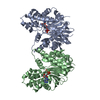
| ||||||||
|---|---|---|---|---|---|---|---|---|---|
| 1 |
| ||||||||
| Unit cell |
|
-Components
| #1: Protein | Mass: 39473.828 Da / Num. of mol.: 2 Source method: isolated from a genetically manipulated source Details: GAGAG expresion tag in N-terminal. These five aminoacids should be renumbered from -4 to 0. Source: (gene. exp.) Pseudomonas aeruginosa (bacteria) / Gene: anmK, PA0666 / Production host: References: UniProt: Q9I5Q5, anhydro-N-acetylmuramic acid kinase #2: Chemical |   Mass: 506.196 Da / Num. of mol.: 2 / Source method: obtained synthetically / Formula: C10H17N6O12P3 / Feature type: SUBJECT OF INVESTIGATION / Comment: AMP-PNP, energy-carrying molecule analogue*YM Mass: 506.196 Da / Num. of mol.: 2 / Source method: obtained synthetically / Formula: C10H17N6O12P3 / Feature type: SUBJECT OF INVESTIGATION / Comment: AMP-PNP, energy-carrying molecule analogue*YM#3: Chemical |   Mass: 24.305 Da / Num. of mol.: 2 / Source method: obtained synthetically / Formula: Mg / Feature type: SUBJECT OF INVESTIGATION Mass: 24.305 Da / Num. of mol.: 2 / Source method: obtained synthetically / Formula: Mg / Feature type: SUBJECT OF INVESTIGATION#4: Water | ChemComp-HOH / |  Mass: 18.015 Da / Num. of mol.: 123 / Source method: isolated from a natural source / Formula: H2O Mass: 18.015 Da / Num. of mol.: 123 / Source method: isolated from a natural source / Formula: H2OHas ligand of interest | Y | |
|---|
-Experimental details
-Experiment
| Experiment | Method: X-RAY DIFFRACTION / Number of used crystals: 1 |
|---|
- Sample preparation
| Crystal | Density Matthews: 2.62 Å3/Da / Density % sol: 54.32 % / Description: Hexagonal crystals |
|---|---|
| Crystal grow | Temperature: 291 K / Method: vapor diffusion, sitting drop / Details: 20% PEG 3350, Na/K tartrate 0.2M |
-Data collection
| Diffraction | Mean temperature: 100 K / Serial crystal experiment: N | ||||||||||||||||||
|---|---|---|---|---|---|---|---|---|---|---|---|---|---|---|---|---|---|---|---|
| Diffraction source | Source: SYNCHROTRON / Site: ALBA / Beamline: XALOC / Wavelength: 0.97926 Å | ||||||||||||||||||
| Detector | Type: DECTRIS PILATUS 6M / Detector: PIXEL / Date: Dec 16, 2022 | ||||||||||||||||||
| Radiation | Protocol: SINGLE WAVELENGTH / Monochromatic (M) / Laue (L): M / Scattering type: x-ray | ||||||||||||||||||
| Radiation wavelength | Wavelength: 0.97926 Å / Relative weight: 1 | ||||||||||||||||||
| Reflection twin |
| ||||||||||||||||||
| Reflection | Resolution: 2.2→47.024 Å / Num. obs: 41089 / % possible obs: 100 % / Redundancy: 20.8 % / CC1/2: 0.999 / Rmerge(I) obs: 0.099 / Net I/σ(I): 21 | ||||||||||||||||||
| Reflection shell | Resolution: 2.2→2.27 Å / Redundancy: 21.3 % / Rmerge(I) obs: 1.717 / Mean I/σ(I) obs: 2.2 / Num. unique obs: 3557 / CC1/2: 0.701 / % possible all: 100 |
- Processing
| Software |
| ||||||||||||||||||||||||||||||||||||||||||||||||||||||||||||||||||||||||||||||||||||||||||||||||||||||||||||||||||||||||||||||||||||||||||||||||||||||||||||||||
|---|---|---|---|---|---|---|---|---|---|---|---|---|---|---|---|---|---|---|---|---|---|---|---|---|---|---|---|---|---|---|---|---|---|---|---|---|---|---|---|---|---|---|---|---|---|---|---|---|---|---|---|---|---|---|---|---|---|---|---|---|---|---|---|---|---|---|---|---|---|---|---|---|---|---|---|---|---|---|---|---|---|---|---|---|---|---|---|---|---|---|---|---|---|---|---|---|---|---|---|---|---|---|---|---|---|---|---|---|---|---|---|---|---|---|---|---|---|---|---|---|---|---|---|---|---|---|---|---|---|---|---|---|---|---|---|---|---|---|---|---|---|---|---|---|---|---|---|---|---|---|---|---|---|---|---|---|---|---|---|---|---|
| Refinement | Method to determine structure: MOLECULAR REPLACEMENT / Resolution: 2.2→47.024 Å / Cor.coef. Fo:Fc: 0.964 / Cor.coef. Fo:Fc free: 0.951 / WRfactor Rfree: 0.212 / WRfactor Rwork: 0.172 / SU B: 8.792 / SU ML: 0.126 / Average fsc free: 0.9577 / Average fsc work: 0.9763 / Cross valid method: FREE R-VALUE / ESU R: 0.05 / ESU R Free: 0.041 Details: Hydrogens have been added in their riding positions
| ||||||||||||||||||||||||||||||||||||||||||||||||||||||||||||||||||||||||||||||||||||||||||||||||||||||||||||||||||||||||||||||||||||||||||||||||||||||||||||||||
| Solvent computation | Ion probe radii: 0.8 Å / Shrinkage radii: 0.8 Å / VDW probe radii: 1.2 Å / Solvent model: MASK BULK SOLVENT | ||||||||||||||||||||||||||||||||||||||||||||||||||||||||||||||||||||||||||||||||||||||||||||||||||||||||||||||||||||||||||||||||||||||||||||||||||||||||||||||||
| Displacement parameters | Biso mean: 53.71 Å2
| ||||||||||||||||||||||||||||||||||||||||||||||||||||||||||||||||||||||||||||||||||||||||||||||||||||||||||||||||||||||||||||||||||||||||||||||||||||||||||||||||
| Refinement step | Cycle: LAST / Resolution: 2.2→47.024 Å
| ||||||||||||||||||||||||||||||||||||||||||||||||||||||||||||||||||||||||||||||||||||||||||||||||||||||||||||||||||||||||||||||||||||||||||||||||||||||||||||||||
| Refine LS restraints |
| ||||||||||||||||||||||||||||||||||||||||||||||||||||||||||||||||||||||||||||||||||||||||||||||||||||||||||||||||||||||||||||||||||||||||||||||||||||||||||||||||
| LS refinement shell |
| ||||||||||||||||||||||||||||||||||||||||||||||||||||||||||||||||||||||||||||||||||||||||||||||||||||||||||||||||||||||||||||||||||||||||||||||||||||||||||||||||
| Refinement TLS params. | Method: refined / Refine-ID: X-RAY DIFFRACTION
| ||||||||||||||||||||||||||||||||||||||||||||||||||||||||||||||||||||||||||||||||||||||||||||||||||||||||||||||||||||||||||||||||||||||||||||||||||||||||||||||||
| Refinement TLS group |
|
