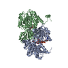
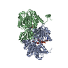
Entry Database : PDB / ID : 8codTitle Crystal structure of S-adenosyl-L-homocysteine hydrolase from Mus musculus in complex with inosine Adenosylhomocysteinase Keywords / / / Function / homology Function Domain/homology Component
/ / / / / / / / / / / / / / / / / / / / / / / / / / / / / / / / / / / / / Biological species Mus musculus (house mouse)Method / / / Resolution : 2.48 Å Authors Saleem-Batcha, R. / Popadic, D. / Koeppl, L.H. / Andexer, J.N. Funding support Organization Grant number Country German Research Foundation (DFG) 235777276/RTG1976 European Research Council (ERC) ERC project 716966 European Union
Journal : Commun Biol / Year : 2024Title : Structure, function and substrate preferences of archaeal S-adenosyl-L-homocysteine hydrolases.Authors : Koeppl, L.H. / Popadic, D. / Saleem-Batcha, R. / Germer, P. / Andexer, J.N. History Deposition Feb 27, 2023 Deposition site / Processing site Revision 1.0 Mar 6, 2024 Provider / Type Revision 1.1 Sep 18, 2024 Group / Category / citation_authorItem _citation.country / _citation.journal_abbrev ... _citation.country / _citation.journal_abbrev / _citation.journal_id_CSD / _citation.journal_id_ISSN / _citation.journal_volume / _citation.page_first / _citation.page_last / _citation.pdbx_database_id_DOI / _citation.pdbx_database_id_PubMed / _citation.title / _citation.year
Show all Show less
 Movie
Movie Controller

 Yorodumi
 Open data
 Basic information
 Components
 Keywords
 Function and homology information
 X-RAY DIFFRACTION /
  Authors
 Germany, European Union, 2items
  Citation
 Structure visualization
 Downloads & links
 Other downloads
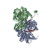
 PDBj
PDBj Assembly
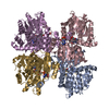
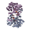
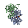


 Mass: 663.425 Da / Num. of mol.: 2 / Source method: obtained synthetically / Formula: C21H27N7O14P2 / Feature type: SUBJECT OF INVESTIGATION / Comment: NAD*YM
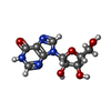
 Mass: 268.226 Da / Num. of mol.: 2 / Source method: obtained synthetically / Formula: C10H12N4O5 / Feature type: SUBJECT OF INVESTIGATION
 Mass: 22.990 Da / Num. of mol.: 2 / Source method: obtained synthetically / Formula: Na
 Mass: 18.015 Da / Num. of mol.: 322 / Source method: isolated from a natural source / Formula: H2O
 Sample preparation
 / Beamline: X06SA / Wavelength: 1 Å
 Processing