[English] 日本語
 Yorodumi
- PDB-8cmu: High resolution structure of the coagulation Factor XIII A2B2 het... -
+ Open data
- Basic information
| Entry | Database: PDB / ID: 8cmu | |||||||||
|---|---|---|---|---|---|---|---|---|---|---|
| Title | High resolution structure of the coagulation Factor XIII A2B2 heterotetramer complex. | |||||||||
 Components Components |
| |||||||||
 Keywords Keywords | BLOOD CLOTTING / Factor XIII | |||||||||
| Function / homology |  Function and homology information Function and homology informationprotein-glutamine gamma-glutamyltransferase / protein-glutamine gamma-glutamyltransferase activity / transferase complex / peptide cross-linking / blood coagulation, fibrin clot formation / Common Pathway of Fibrin Clot Formation / platelet alpha granule lumen / : / blood coagulation / Platelet degranulation ...protein-glutamine gamma-glutamyltransferase / protein-glutamine gamma-glutamyltransferase activity / transferase complex / peptide cross-linking / blood coagulation, fibrin clot formation / Common Pathway of Fibrin Clot Formation / platelet alpha granule lumen / : / blood coagulation / Platelet degranulation / Interleukin-4 and Interleukin-13 signaling / blood microparticle / extracellular space / extracellular region / metal ion binding Similarity search - Function | |||||||||
| Biological species |  Homo sapiens (human) Homo sapiens (human) | |||||||||
| Method | ELECTRON MICROSCOPY / single particle reconstruction / cryo EM / Resolution: 2.41 Å | |||||||||
 Authors Authors | Singh, S. / Urgular, D. / Hagelueken, G. / Geyer, M. / Biswas, A. | |||||||||
| Funding support |  Germany, 2items Germany, 2items
| |||||||||
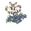
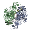
 Citation Citation |  Journal: Blood / Year: 2025 Journal: Blood / Year: 2025Title: Cryo-EM structure of the human native plasma coagulation factor XIII complex. Authors: Sneha Singh / Gregor Hagelueken / Deniz Ugurlar / Samhitha Urs Ramaraje Urs / Amit Sharma / Manoranjan Mahapatra / Friedel Drepper / Diana Imhof / Pitter F Huesgen / Johannes Oldenburg / ...Authors: Sneha Singh / Gregor Hagelueken / Deniz Ugurlar / Samhitha Urs Ramaraje Urs / Amit Sharma / Manoranjan Mahapatra / Friedel Drepper / Diana Imhof / Pitter F Huesgen / Johannes Oldenburg / Matthias Geyer / Arijit Biswas /    Abstract: The structure of human coagulation factor XIII (FXIII), a heterotetrameric plasma protransglutaminase that covalently cross-links preformed fibrin polymers, remains elusive until today. The ...The structure of human coagulation factor XIII (FXIII), a heterotetrameric plasma protransglutaminase that covalently cross-links preformed fibrin polymers, remains elusive until today. The heterotetrameric complex is composed of 2 catalytic FXIII-A and 2 protective FXIII-B subunits. Structural etiology underlying FXIII deficiency has so far been derived from crystallographic structures, all of which are currently available for the FXIII-A2 homodimer only. Here, we present the cryogenic electron microscopy (cryo-EM) structure of a native, human plasma-derived FXIII-A2B2 complex at 2.4 Å resolution. The structure provides detailed information on FXIII subunit interacting interfaces as the 2 subunits interact strongly in plasma. The native FXIII-A2B2 complex reveals a pseudosymmetric heterotetramer of 2 FXIII-B monomers intercalating with a symmetric FXIII-A2 dimer forming a "crown"-like assembly. The symmetry axes of the A2 and B2 homodimers are twisted relative to each other such that Sushi domain 1 interacts with the catalytic core of the A subunit, and Sushi domain 2 with the symmetry related A' subunit, and vice versa. We also report 4 novel mutations in the F13A1 gene encoding the FXIII-A subunit from a cohort of patients with severe FXIII deficiency. Our structure reveals the etiological basis of homozygous and heterozygous pathogenic mutations and explains the conditional dominant negative effects of heterozygous mutations. This atomistic description of complex interfaces is consistent with previous biochemical data and shows a congruence between the structural biochemistry of the FXIII complex and the clinical features of FXIII deficiency. | |||||||||
| History |
|
- Structure visualization
| Structure viewer | Molecule:  Molmil Molmil Jmol/JSmol Jmol/JSmol |
|---|
- Downloads & links
- Download
| PDBx/mmCIF format |  8cmu.cif.gz 8cmu.cif.gz | 458 KB | Display |  PDBx/mmCIF format PDBx/mmCIF format |
|---|---|---|---|---|
| PDB format |  pdb8cmu.ent.gz pdb8cmu.ent.gz | 292.4 KB | Display |  PDB format PDB format |
| PDBx/mmJSON format |  8cmu.json.gz 8cmu.json.gz | Tree view |  PDBx/mmJSON format PDBx/mmJSON format | |
| Others |  Other downloads Other downloads |
-Validation report
| Arichive directory |  https://data.pdbj.org/pub/pdb/validation_reports/cm/8cmu https://data.pdbj.org/pub/pdb/validation_reports/cm/8cmu ftp://data.pdbj.org/pub/pdb/validation_reports/cm/8cmu ftp://data.pdbj.org/pub/pdb/validation_reports/cm/8cmu | HTTPS FTP |
|---|
-Related structure data
| Related structure data |  16746MC  8cmtC M: map data used to model this data C: citing same article ( |
|---|---|
| Similar structure data | Similarity search - Function & homology  F&H Search F&H Search |
- Links
Links
- Assembly
| Deposited unit | 
|
|---|---|
| 1 |
|
- Components
| #1: Protein | Mass: 83365.109 Da / Num. of mol.: 2 / Source method: isolated from a natural source / Source: (natural)  Homo sapiens (human) Homo sapiens (human)References: UniProt: P00488, protein-glutamine gamma-glutamyltransferase #2: Protein | Mass: 75600.594 Da / Num. of mol.: 2 / Source method: isolated from a natural source / Source: (natural)  Homo sapiens (human) / References: UniProt: P05160 Homo sapiens (human) / References: UniProt: P05160#3: Water | ChemComp-HOH / | Has protein modification | Y | |
|---|
-Experimental details
-Experiment
| Experiment | Method: ELECTRON MICROSCOPY |
|---|---|
| EM experiment | Aggregation state: PARTICLE / 3D reconstruction method: single particle reconstruction |
- Sample preparation
| Component | Name: Factor XIII A2B2 heterotetramer / Type: COMPLEX / Entity ID: #1-#2 / Source: NATURAL |
|---|---|
| Source (natural) | Organism:  Homo sapiens (human) Homo sapiens (human) |
| Buffer solution | pH: 7.5 |
| Specimen | Embedding applied: NO / Shadowing applied: NO / Staining applied: NO / Vitrification applied: YES |
| Vitrification | Cryogen name: ETHANE |
- Electron microscopy imaging
| Experimental equipment |  Model: Titan Krios / Image courtesy: FEI Company |
|---|---|
| Microscopy | Model: FEI TITAN KRIOS |
| Electron gun | Electron source:  FIELD EMISSION GUN / Accelerating voltage: 300 kV / Illumination mode: FLOOD BEAM FIELD EMISSION GUN / Accelerating voltage: 300 kV / Illumination mode: FLOOD BEAM |
| Electron lens | Mode: BRIGHT FIELD / Nominal defocus max: 1800 nm / Nominal defocus min: 800 nm |
| Image recording | Electron dose: 50 e/Å2 / Film or detector model: FEI FALCON IV (4k x 4k) |
- Processing
| Software |
| ||||||||||||||||||||||||
|---|---|---|---|---|---|---|---|---|---|---|---|---|---|---|---|---|---|---|---|---|---|---|---|---|---|
| CTF correction | Type: PHASE FLIPPING AND AMPLITUDE CORRECTION | ||||||||||||||||||||||||
| 3D reconstruction | Resolution: 2.41 Å / Resolution method: FSC 0.143 CUT-OFF / Num. of particles: 333272 / Symmetry type: POINT | ||||||||||||||||||||||||
| Refinement | Cross valid method: NONE Stereochemistry target values: GeoStd + Monomer Library + CDL v1.2 | ||||||||||||||||||||||||
| Displacement parameters | Biso mean: 51 Å2 | ||||||||||||||||||||||||
| Refine LS restraints |
|
 Movie
Movie Controller


 PDBj
PDBj


