 Movie
Movie Controller

[English] 日本語
 Yorodumi
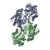
- PDB-8cek: Succinyl-CoA Reductase from Clostridium kluyveri (SucD) with NADPH -
+ Open data

- Basic information
| Entry | Database: PDB / ID: 8cek | ||||||
|---|---|---|---|---|---|---|---|
| Title | Succinyl-CoA Reductase from Clostridium kluyveri (SucD) with NADPH | ||||||
 Components Components | Succinate-semialdehyde dehydrogenase (acetylating) | ||||||
 Keywords Keywords | OXIDOREDUCTASE / SSA / succinic semialdehyde / NADPH / NADP+ / SucD / ssr / succinyl-CoA / mesaconyl-CoA / mesaconyl-C1-CoA / SucD_Ck / CkSucD | ||||||
| Function / homology | succinate-semialdehyde dehydrogenase (acylating) / oxidoreductase activity, acting on the aldehyde or oxo group of donors, NAD or NADP as acceptor / Aldehyde dehydrogenase domain / Aldehyde dehydrogenase family / Aldehyde dehydrogenase, C-terminal / Aldehyde dehydrogenase, N-terminal / Aldehyde/histidinol dehydrogenase / NADP NICOTINAMIDE-ADENINE-DINUCLEOTIDE PHOSPHATE / Succinate-semialdehyde dehydrogenase (acetylating) Function and homology information Function and homology information | ||||||
| Biological species |  Clostridium kluyveri (bacteria) Clostridium kluyveri (bacteria) | ||||||
| Method |  X-RAY DIFFRACTION / SYNCHROTRON / MOLECULAR REPLACEMENT / Resolution: 2.15 Å X-RAY DIFFRACTION / SYNCHROTRON / MOLECULAR REPLACEMENT / Resolution: 2.15 Å | ||||||
 Authors Authors | Pfister, P. / Diehl, C. / Erb, T.J. | ||||||
| Funding support |  Germany, 1items Germany, 1items
| ||||||
 Citation Citation | Journal: Biochemistry / Year: 2023 Title: Enhancing the Substrate Specificity of Clostridium Succinyl-CoA Reductase for Synthetic Biology and Biocatalysis. Authors: Pfister, P. / Diehl, C. / Hammarlund, E. / Carrillo, M. / Erb, T.J. | ||||||
| History |
|
- Structure visualization
| Structure viewer | Molecule: MolmilJmol/JSmol |
|---|
- Downloads & links
-Download
| PDBx/mmCIF format | 8cek.cif.gz | 707.6 KB | Display | PDBx/mmCIF format |
|---|---|---|---|---|
| PDB format | pdb8cek.ent.gz | 591.4 KB | Display | PDB format |
| PDBx/mmJSON format | 8cek.json.gz | Tree view | PDBx/mmJSON format | |
| Others |  Other downloads Other downloads |
-Validation report
| Arichive directory | https://data.pdbj.org/pub/pdb/validation_reports/ce/8cekftp://data.pdbj.org/pub/pdb/validation_reports/ce/8cek | HTTPS FTP |
|---|
-Related structure data

-Links
 PDBj
PDBj

- Assembly
| Deposited unit | 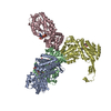
| ||||||||
|---|---|---|---|---|---|---|---|---|---|
| 1 | 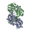
| ||||||||
| 2 | 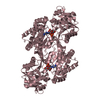
| ||||||||
| 3 | 
| ||||||||
| Unit cell |
| ||||||||
| Components on special symmetry positions |
|
-Components
| #1: Protein | Mass: 49029.910 Da / Num. of mol.: 4 Source method: isolated from a genetically manipulated source Source: (gene. exp.) Clostridium kluyveri (bacteria) / Gene: sucD, CKL_3015 / Production host: References: UniProt: P38947, succinate-semialdehyde dehydrogenase (acylating) #2: Chemical | ChemComp-NAP /   Mass: 743.405 Da / Num. of mol.: 4 / Source method: obtained synthetically / Formula: C21H28N7O17P3 / Feature type: SUBJECT OF INVESTIGATION Mass: 743.405 Da / Num. of mol.: 4 / Source method: obtained synthetically / Formula: C21H28N7O17P3 / Feature type: SUBJECT OF INVESTIGATION#3: Water | ChemComp-HOH / |  Mass: 18.015 Da / Num. of mol.: 1066 / Source method: isolated from a natural source / Formula: H2O Mass: 18.015 Da / Num. of mol.: 1066 / Source method: isolated from a natural source / Formula: H2OHas ligand of interest | Y | |
|---|
-Experimental details
-Experiment
| Experiment | Method: X-RAY DIFFRACTION / Number of used crystals: 1 |
|---|
- Sample preparation
| Crystal | Density Matthews: 3.23 Å3/Da / Density % sol: 61.87 % |
|---|---|
| Crystal grow | Temperature: 288.15 K / Method: vapor diffusion, sitting drop / pH: 6.5 Details: 45 % w/v Pentaerythritol Propoxylate (5/4 PO/OH) 100 mM MES; pH 6.5 400 mM Potassium chloride PH range: 6.5-7.8 |
-Data collection
| Diffraction | Mean temperature: 80 K / Serial crystal experiment: N |
|---|---|
| Diffraction source | Source: SYNCHROTRON / Site: PETRA III, EMBL c/o DESY / Beamline: P13 (MX1) / Wavelength: 0.9762 Å |
| Detector | Type: DECTRIS PILATUS 6M / Detector: PIXEL / Date: Mar 30, 2019 |
| Radiation | Protocol: SINGLE WAVELENGTH / Monochromatic (M) / Laue (L): M / Scattering type: x-ray |
| Radiation wavelength | Wavelength: 0.9762 Å / Relative weight: 1 |
| Reflection | Resolution: 2.04→39.44 Å / Num. obs: 159239 / % possible obs: 98.6 % / Redundancy: 12.8 % / CC1/2: 0.998 / Rpim(I) all: 0.059 / Rrim(I) all: 0.215 / Net I/σ(I): 8.5 / Num. measured all: 2045343 |
| Reflection shell | Resolution: 2.04→2.15 Å / % possible obs: 90.4 % / Redundancy: 8 % / Num. measured all: 169254 / Num. unique obs: 21156 / CC1/2: 0.329 / Rpim(I) all: 0.966 / Rrim(I) all: 2.931 / Net I/σ(I) obs: 0.6 |
- Processing
| Software |
| |||||||||||||||||||||||||||||||||||||||||||||||||||||||||||||||||||||||||||||||||||||||||||||||||||||||||
|---|---|---|---|---|---|---|---|---|---|---|---|---|---|---|---|---|---|---|---|---|---|---|---|---|---|---|---|---|---|---|---|---|---|---|---|---|---|---|---|---|---|---|---|---|---|---|---|---|---|---|---|---|---|---|---|---|---|---|---|---|---|---|---|---|---|---|---|---|---|---|---|---|---|---|---|---|---|---|---|---|---|---|---|---|---|---|---|---|---|---|---|---|---|---|---|---|---|---|---|---|---|---|---|---|---|---|
| Refinement | Method to determine structure: MOLECULAR REPLACEMENT / Resolution: 2.15→29.66 Å / SU ML: 0.25 / Cross valid method: FREE R-VALUE / σ(F): 1.34 / Phase error: 25.84 / Stereochemistry target values: ML
| |||||||||||||||||||||||||||||||||||||||||||||||||||||||||||||||||||||||||||||||||||||||||||||||||||||||||
| Solvent computation | Shrinkage radii: 0.9 Å / VDW probe radii: 1.1 Å / Solvent model: FLAT BULK SOLVENT MODEL | |||||||||||||||||||||||||||||||||||||||||||||||||||||||||||||||||||||||||||||||||||||||||||||||||||||||||
| Refinement step | Cycle: LAST / Resolution: 2.15→29.66 Å
| |||||||||||||||||||||||||||||||||||||||||||||||||||||||||||||||||||||||||||||||||||||||||||||||||||||||||
| Refine LS restraints |
| |||||||||||||||||||||||||||||||||||||||||||||||||||||||||||||||||||||||||||||||||||||||||||||||||||||||||
| LS refinement shell |
| |||||||||||||||||||||||||||||||||||||||||||||||||||||||||||||||||||||||||||||||||||||||||||||||||||||||||
| Refinement TLS params. | Method: refined / Origin x: 14.3316 Å / Origin y: -23.6325 Å / Origin z: 47.3039 Å
| |||||||||||||||||||||||||||||||||||||||||||||||||||||||||||||||||||||||||||||||||||||||||||||||||||||||||
| Refinement TLS group | Selection details: all |
