 Movie
Movie Controller

[English] 日本語
 Yorodumi
- PDB-8ce3: Crystal structure of MGAT5 (alpha-1,6-mannosylglycoprotein 6-beta... -
+ Open data

- Basic information
| Entry | Database: PDB / ID: 8ce3 | ||||||||||||
|---|---|---|---|---|---|---|---|---|---|---|---|---|---|
| Title | Crystal structure of MGAT5 (alpha-1,6-mannosylglycoprotein 6-beta-N-acetylglucosaminyltransferase V) luminal domain with a Lys329-Ile345 loop truncation, in complex with 3D fragment 2548 | ||||||||||||
 Components Components | Alpha-1,6-mannosylglycoprotein 6-beta-N-acetylglucosaminyltransferase A | ||||||||||||
 Keywords Keywords | CARBOHYDRATE / Glycosyltransferase / fragment | ||||||||||||
| Function / homology |  Function and homology information Function and homology informationalpha-1,6-mannosyl-glycoprotein 6-beta-N-acetylglucosaminyltransferase / alpha-1,6-mannosylglycoprotein 6-beta-N-acetylglucosaminyltransferase activity / N-Glycan antennae elongation / positive regulation of receptor signaling pathway via STAT / : / protein N-linked glycosylation / protein phosphatase inhibitor activity / manganese ion binding / Maturation of spike protein / positive regulation of cell migration ...alpha-1,6-mannosyl-glycoprotein 6-beta-N-acetylglucosaminyltransferase / alpha-1,6-mannosylglycoprotein 6-beta-N-acetylglucosaminyltransferase activity / N-Glycan antennae elongation / positive regulation of receptor signaling pathway via STAT / : / protein N-linked glycosylation / protein phosphatase inhibitor activity / manganese ion binding / Maturation of spike protein / positive regulation of cell migration / viral protein processing / Golgi membrane / Golgi apparatus / extracellular exosome / membrane Similarity search - Function | ||||||||||||
| Biological species |  Homo sapiens (human) Homo sapiens (human) | ||||||||||||
| Method |  X-RAY DIFFRACTION / SYNCHROTRON / MOLECULAR REPLACEMENT / Resolution: 1.89 Å X-RAY DIFFRACTION / SYNCHROTRON / MOLECULAR REPLACEMENT / Resolution: 1.89 Å | ||||||||||||
 Authors Authors | Gilio, A.K. / Darby, J.F. / Wu, L. / Davies, G.J. | ||||||||||||
| Funding support |  United Kingdom, European Union, 3items United Kingdom, European Union, 3items
| ||||||||||||
 Citation Citation | Journal: Chemrxiv / Year: 2025 Title: Design, Modular Synthesis and Screening of 58 Shape-Diverse 3-D Fragments Authors: Downes, T.D. / Jones, S.P. / Firth, J.D. / Darby, J.F. / Gilio, A.K. / Klein, H.F. / Blakemore, D.C. / de Fusco, C. / Roughley, S.D. / Vidler, L.R. / Whatton, M.A. / Woolford, A.J.A. / ...Authors: Downes, T.D. / Jones, S.P. / Firth, J.D. / Darby, J.F. / Gilio, A.K. / Klein, H.F. / Blakemore, D.C. / de Fusco, C. / Roughley, S.D. / Vidler, L.R. / Whatton, M.A. / Woolford, A.J.A. / Wrigley, G.L. / Hubbard, R.E. / Wu, L. / Davies, G.J. / O'Brien, P. | ||||||||||||
| History |
|
- Structure visualization
| Structure viewer | Molecule: MolmilJmol/JSmol |
|---|
- Downloads & links
-Download
| PDBx/mmCIF format | 8ce3.cif.gz | 232.1 KB | Display | PDBx/mmCIF format |
|---|---|---|---|---|
| PDB format | pdb8ce3.ent.gz | 176.2 KB | Display | PDB format |
| PDBx/mmJSON format | 8ce3.json.gz | Tree view | PDBx/mmJSON format | |
| Others |  Other downloads Other downloads |
-Validation report
| Summary document | 8ce3_validation.pdf.gz | 807.5 KB | Display | wwPDB validaton report |
|---|---|---|---|---|
| Full document | 8ce3_full_validation.pdf.gz | 814.7 KB | Display | |
| Data in XML | 8ce3_validation.xml.gz | 39.5 KB | Display | |
| Data in CIF | 8ce3_validation.cif.gz | 57.1 KB | Display | |
| Arichive directory | https://data.pdbj.org/pub/pdb/validation_reports/ce/8ce3ftp://data.pdbj.org/pub/pdb/validation_reports/ce/8ce3 | HTTPS FTP |
-Related structure data
| Similar structure data |
|---|
-Links
 PDBj
PDBj- Assembly
| Deposited unit | 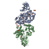
| ||||||||||||
|---|---|---|---|---|---|---|---|---|---|---|---|---|---|
| 1 |
| ||||||||||||
| Unit cell |
| ||||||||||||
| Noncrystallographic symmetry (NCS) | NCS domain:
NCS domain segments: Component-ID: 1 / Ens-ID: 1 / Beg auth comp-ID: SER / Beg label comp-ID: SER / End auth comp-ID: LEU / End label comp-ID: LEU / Refine code: 1 / Auth asym-ID: A / Label asym-ID: A / Auth seq-ID: 214 - 728 / Label seq-ID: 1 - 515
NCS ensembles : (Details: Local NCS retraints between domains: 1 2) |
-Components
| #1: Protein | Mass: 58996.906 Da / Num. of mol.: 2 Source method: isolated from a genetically manipulated source Source: (gene. exp.) Homo sapiens (human) / Gene: MGAT5, GGNT5 / Production host:  Trichoplusia ni (cabbage looper) Trichoplusia ni (cabbage looper)References: UniProt: Q09328, alpha-1,6-mannosyl-glycoprotein 6-beta-N-acetylglucosaminyltransferase #2: Chemical | ChemComp-EDO /   Mass: 62.068 Da / Num. of mol.: 7 / Source method: obtained synthetically / Formula: C2H6O2 Mass: 62.068 Da / Num. of mol.: 7 / Source method: obtained synthetically / Formula: C2H6O2#3: Chemical | ChemComp-UE0 / ( | Mass: 261.316 Da / Num. of mol.: 1 / Source method: obtained synthetically / Formula: C15H19NO3 / Feature type: SUBJECT OF INVESTIGATION #4: Chemical | ChemComp-SO4 /   Mass: 96.063 Da / Num. of mol.: 13 / Source method: obtained synthetically / Formula: SO4 Mass: 96.063 Da / Num. of mol.: 13 / Source method: obtained synthetically / Formula: SO4#5: Water | ChemComp-HOH / |  Mass: 18.015 Da / Num. of mol.: 349 / Source method: isolated from a natural source / Formula: H2O Mass: 18.015 Da / Num. of mol.: 349 / Source method: isolated from a natural source / Formula: H2OHas ligand of interest | Y | Has protein modification | Y | |
|---|
-Experimental details
-Experiment
| Experiment | Method: X-RAY DIFFRACTION / Number of used crystals: 1 |
|---|
- Sample preparation
| Crystal | Density Matthews: 2.25 Å3/Da / Density % sol: 45.27 % |
|---|---|
| Crystal grow | Temperature: 293 K / Method: vapor diffusion, sitting drop Details: 0.1 M HEPES pH 8.0, 0.3 M Li2SO4, 30 % (w/v) PEG 3350, 10 % (v/v) ethylene glycol |
-Data collection
| Diffraction | Mean temperature: 100 K / Serial crystal experiment: N |
|---|---|
| Diffraction source | Source: SYNCHROTRON / Site: Diamond / Beamline: I04 / Wavelength: 0.95 Å |
| Detector | Type: DECTRIS EIGER X 16M / Detector: PIXEL / Date: Mar 3, 2019 |
| Radiation | Protocol: SINGLE WAVELENGTH / Monochromatic (M) / Laue (L): M / Scattering type: x-ray |
| Radiation wavelength | Wavelength: 0.95 Å / Relative weight: 1 |
| Reflection | Resolution: 1.89→60.47 Å / Num. obs: 80017 / % possible obs: 97.26 % / Redundancy: 3.5 % / CC1/2: 1 / Rrim(I) all: 0.047 / Net I/σ(I): 13.6 |
| Reflection shell | Resolution: 1.89→1.92 Å / Num. unique obs: 5618 / CC1/2: 0.6 / Rrim(I) all: 1.297 |
- Processing
| Software |
| |||||||||||||||||||||||||||||||||||||||||||||||||||||||||||||||||||||||||||||||||||||||||||||||||||||||||||||||||||||||||||||||||||||||||||||||||||||||||||||||||||||||||||||||||||||||||||||||||||||||||||||||||||||||||||||||||||||||
|---|---|---|---|---|---|---|---|---|---|---|---|---|---|---|---|---|---|---|---|---|---|---|---|---|---|---|---|---|---|---|---|---|---|---|---|---|---|---|---|---|---|---|---|---|---|---|---|---|---|---|---|---|---|---|---|---|---|---|---|---|---|---|---|---|---|---|---|---|---|---|---|---|---|---|---|---|---|---|---|---|---|---|---|---|---|---|---|---|---|---|---|---|---|---|---|---|---|---|---|---|---|---|---|---|---|---|---|---|---|---|---|---|---|---|---|---|---|---|---|---|---|---|---|---|---|---|---|---|---|---|---|---|---|---|---|---|---|---|---|---|---|---|---|---|---|---|---|---|---|---|---|---|---|---|---|---|---|---|---|---|---|---|---|---|---|---|---|---|---|---|---|---|---|---|---|---|---|---|---|---|---|---|---|---|---|---|---|---|---|---|---|---|---|---|---|---|---|---|---|---|---|---|---|---|---|---|---|---|---|---|---|---|---|---|---|---|---|---|---|---|---|---|---|---|---|---|---|---|---|---|---|---|
| Refinement | Method to determine structure: MOLECULAR REPLACEMENT / Resolution: 1.89→60.465 Å / Cor.coef. Fo:Fc: 0.969 / Cor.coef. Fo:Fc free: 0.952 / WRfactor Rfree: 0.241 / WRfactor Rwork: 0.197 / SU B: 4.945 / SU ML: 0.137 / Average fsc free: 0.9498 / Average fsc work: 0.9652 / Cross valid method: FREE R-VALUE / ESU R: 0.165 / ESU R Free: 0.151 Details: Hydrogens have been added in their riding positions
| |||||||||||||||||||||||||||||||||||||||||||||||||||||||||||||||||||||||||||||||||||||||||||||||||||||||||||||||||||||||||||||||||||||||||||||||||||||||||||||||||||||||||||||||||||||||||||||||||||||||||||||||||||||||||||||||||||||||
| Solvent computation | Ion probe radii: 0.8 Å / Shrinkage radii: 0.8 Å / VDW probe radii: 1.2 Å / Solvent model: MASK BULK SOLVENT | |||||||||||||||||||||||||||||||||||||||||||||||||||||||||||||||||||||||||||||||||||||||||||||||||||||||||||||||||||||||||||||||||||||||||||||||||||||||||||||||||||||||||||||||||||||||||||||||||||||||||||||||||||||||||||||||||||||||
| Displacement parameters | Biso mean: 54.819 Å2
| |||||||||||||||||||||||||||||||||||||||||||||||||||||||||||||||||||||||||||||||||||||||||||||||||||||||||||||||||||||||||||||||||||||||||||||||||||||||||||||||||||||||||||||||||||||||||||||||||||||||||||||||||||||||||||||||||||||||
| Refinement step | Cycle: LAST / Resolution: 1.89→60.465 Å
| |||||||||||||||||||||||||||||||||||||||||||||||||||||||||||||||||||||||||||||||||||||||||||||||||||||||||||||||||||||||||||||||||||||||||||||||||||||||||||||||||||||||||||||||||||||||||||||||||||||||||||||||||||||||||||||||||||||||
| Refine LS restraints |
| |||||||||||||||||||||||||||||||||||||||||||||||||||||||||||||||||||||||||||||||||||||||||||||||||||||||||||||||||||||||||||||||||||||||||||||||||||||||||||||||||||||||||||||||||||||||||||||||||||||||||||||||||||||||||||||||||||||||
| Refine LS restraints NCS |
| |||||||||||||||||||||||||||||||||||||||||||||||||||||||||||||||||||||||||||||||||||||||||||||||||||||||||||||||||||||||||||||||||||||||||||||||||||||||||||||||||||||||||||||||||||||||||||||||||||||||||||||||||||||||||||||||||||||||
| LS refinement shell | Refine-ID: X-RAY DIFFRACTION / Total num. of bins used: 20
|
