 Movie
Movie Controller

[English] 日本語
 Yorodumi
- PDB-8ccz: Crystal structure of human Sirt3 in complex with an inhibiting HI... -
+ Open data

- Basic information
| Entry | Database: PDB / ID: 8ccz | ||||||
|---|---|---|---|---|---|---|---|
| Title | Crystal structure of human Sirt3 in complex with an inhibiting HIV1 Tat-37-59 peptide | ||||||
 Components Components |
| ||||||
 Keywords Keywords | SIGNALING PROTEIN / Sirtuin / inhibitor / HIV / complex | ||||||
| Function / homology |  Function and homology information Function and homology informationviral gene expression / trans-activation response element binding / regulatory region RNA binding / positive regulation of superoxide dismutase activity / positive regulation of catalase activity / protein serine/threonine phosphatase inhibitor activity / NAD-dependent protein lysine delactylase activity / positive regulation of ceramide biosynthetic process / positive regulation of viral transcription / symbiont-mediated perturbation of host chromatin organization ...viral gene expression / trans-activation response element binding / regulatory region RNA binding / positive regulation of superoxide dismutase activity / positive regulation of catalase activity / protein serine/threonine phosphatase inhibitor activity / NAD-dependent protein lysine delactylase activity / positive regulation of ceramide biosynthetic process / positive regulation of viral transcription / symbiont-mediated perturbation of host chromatin organization / Maturation of TCA enzymes and regulation of TCA cycle / peptidyl-lysine deacetylation / symbiont-mediated suppression of host translation initiation / NAD-dependent protein lysine deacetylase activity / host cell nucleolus / protein acetyllysine N-acetyltransferase / protein deacetylation / histone deacetylase activity, NAD-dependent / positive regulation of oxidative phosphorylation / actinin binding / Regulation of FOXO transcriptional activity by acetylation / protein lysine deacetylase activity / cellular response to stress / negative regulation of reactive oxygen species metabolic process / NAD+ binding / Mitochondrial unfolded protein response (UPRmt) / FOXO-mediated transcription of oxidative stress, metabolic and neuronal genes / RNA-binding transcription regulator activity / Transferases; Acyltransferases; Transferring groups other than aminoacyl groups / cyclin binding / aerobic respiration / positive regulation of transcription elongation by RNA polymerase II / Transcriptional activation of mitochondrial biogenesis / positive regulation of insulin secretion / negative regulation of ERK1 and ERK2 cascade / sequence-specific DNA binding / host cell cytoplasm / symbiont-mediated suppression of host innate immune response / symbiont-mediated suppression of host type I interferon-mediated signaling pathway / mitochondrial matrix / protein domain specific binding / DNA-templated transcription / enzyme binding / protein-containing complex / mitochondrion / extracellular region / zinc ion binding / nucleoplasm / metal ion binding / nucleus Similarity search - Function | ||||||
| Biological species |  Homo sapiens (human) Homo sapiens (human)  Human immunodeficiency virus 1 Human immunodeficiency virus 1 | ||||||
| Method |  X-RAY DIFFRACTION / SYNCHROTRON / MOLECULAR REPLACEMENT / Resolution: 1.95 Å X-RAY DIFFRACTION / SYNCHROTRON / MOLECULAR REPLACEMENT / Resolution: 1.95 Å | ||||||
 Authors Authors | Adolph, R.S. / Steegborn, C. | ||||||
| Funding support |  Germany, 1items Germany, 1items
| ||||||
 Citation Citation | Journal: Life / Year: 2023 Title: Molecular Mechanism of Sirtuin 1 Inhibition by Human Immunodeficiency Virus 1 Tat Protein. Authors: Adolph, R.S. / Beck, E. / Schweimer, K. / Di Fonzo, A. / Weyand, M. / Rosch, P. / Wohrl, B.M. / Steegborn, C. | ||||||
| History |
|
- Structure visualization
| Structure viewer | Molecule: MolmilJmol/JSmol |
|---|
- Downloads & links
-Download
| PDBx/mmCIF format | 8ccz.cif.gz | 168 KB | Display | PDBx/mmCIF format |
|---|---|---|---|---|
| PDB format | pdb8ccz.ent.gz | 106 KB | Display | PDB format |
| PDBx/mmJSON format | 8ccz.json.gz | Tree view | PDBx/mmJSON format | |
| Others |  Other downloads Other downloads |
-Validation report
| Arichive directory | https://data.pdbj.org/pub/pdb/validation_reports/cc/8cczftp://data.pdbj.org/pub/pdb/validation_reports/cc/8ccz | HTTPS FTP |
|---|
-Related structure data
| Related structure data |  8ccwC 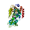 4bvbS S: Starting model for refinement C: citing same article ( |
|---|---|
| Similar structure data |
-Links
 PDBj
PDBj


- Assembly
| Deposited unit | 
| ||||||||||||
|---|---|---|---|---|---|---|---|---|---|---|---|---|---|
| 1 | 
| ||||||||||||
| 2 | 
| ||||||||||||
| Unit cell |
|
-Components
| #1: Protein | Mass: 31340.084 Da / Num. of mol.: 2 Source method: isolated from a genetically manipulated source Source: (gene. exp.) Homo sapiens (human) / Gene: SIRT3, SIR2L3 / Production host:  References: UniProt: Q9NTG7, protein acetyllysine N-acetyltransferase #2: Protein/peptide | Mass: 2741.274 Da / Num. of mol.: 2 / Mutation: C37A / Source method: obtained synthetically / Details: C37A / Source: (synth.) Human immunodeficiency virus 1 / References: UniProt: P12506#3: Chemical |   Mass: 65.409 Da / Num. of mol.: 2 / Source method: obtained synthetically / Formula: Zn Mass: 65.409 Da / Num. of mol.: 2 / Source method: obtained synthetically / Formula: Zn#4: Water | ChemComp-HOH / |  Mass: 18.015 Da / Num. of mol.: 472 / Source method: isolated from a natural source / Formula: H2O Mass: 18.015 Da / Num. of mol.: 472 / Source method: isolated from a natural source / Formula: H2OHas ligand of interest | N | |
|---|
-Experimental details
-Experiment
| Experiment | Method: X-RAY DIFFRACTION / Number of used crystals: 1 |
|---|
- Sample preparation
| Crystal | Density Matthews: 2.38 Å3/Da / Density % sol: 48.32 % / Description: Plates |
|---|---|
| Crystal grow | Temperature: 293.15 K / Method: vapor diffusion, sitting drop Details: 10 mg/ml human Sirt3-(118-399) in 20 mM Tris/HCl, pH 8.0, 150 mM NaCl, 5% (v/v) glycerol, 1 mM TCEP were incubated with 2 mM Tat-37-59 in 10% (v/v) DMSO for 60 min at 293.15 K. The complex ...Details: 10 mg/ml human Sirt3-(118-399) in 20 mM Tris/HCl, pH 8.0, 150 mM NaCl, 5% (v/v) glycerol, 1 mM TCEP were incubated with 2 mM Tat-37-59 in 10% (v/v) DMSO for 60 min at 293.15 K. The complex was crystallized using the sitting-drop vapor-diffusion method at 293.15 K with 100 mM CHES, pH 9.0, 20% (w/v) PEG 8000 as reservoir solution. |
-Data collection
| Diffraction | Mean temperature: 100 K / Serial crystal experiment: N |
|---|---|
| Diffraction source | Source: SYNCHROTRON / Site: BESSY / Beamline: 14.1 / Wavelength: 0.9184 Å |
| Detector | Type: DECTRIS PILATUS 6M / Detector: PIXEL / Date: Jun 13, 2018 |
| Radiation | Monochromator: DCM Si(111) / Protocol: SINGLE WAVELENGTH / Monochromatic (M) / Laue (L): M / Scattering type: x-ray |
| Radiation wavelength | Wavelength: 0.9184 Å / Relative weight: 1 |
| Reflection | Resolution: 1.95→54.24 Å / Num. obs: 46174 / % possible obs: 98.83 % / Redundancy: 5.5 % / Biso Wilson estimate: 30.86 Å2 / CC1/2: 0.949 / CC star: 0.987 / Net I/σ(I): 11.97 |
| Reflection shell | Resolution: 1.95→2.02 Å / Redundancy: 5.5 % / Mean I/σ(I) obs: 0.55 / Num. unique obs: 4615 / CC1/2: 0.445 / CC star: 0.785 / % possible all: 98.88 |
- Processing
| Software |
| ||||||||||||||||||||||||||||||||||||||||||||||||||||||||||||||||||||||||||||||||||||||||||||||||||||||||||||||||||||||||||||||
|---|---|---|---|---|---|---|---|---|---|---|---|---|---|---|---|---|---|---|---|---|---|---|---|---|---|---|---|---|---|---|---|---|---|---|---|---|---|---|---|---|---|---|---|---|---|---|---|---|---|---|---|---|---|---|---|---|---|---|---|---|---|---|---|---|---|---|---|---|---|---|---|---|---|---|---|---|---|---|---|---|---|---|---|---|---|---|---|---|---|---|---|---|---|---|---|---|---|---|---|---|---|---|---|---|---|---|---|---|---|---|---|---|---|---|---|---|---|---|---|---|---|---|---|---|---|---|---|
| Refinement | Method to determine structure: MOLECULAR REPLACEMENT Starting model: 4BVB Resolution: 1.95→54.24 Å / SU ML: 0.2762 / Cross valid method: FREE R-VALUE / σ(F): 1.34 / Phase error: 29.4121 Stereochemistry target values: GeoStd + Monomer Library + CDL v1.2
| ||||||||||||||||||||||||||||||||||||||||||||||||||||||||||||||||||||||||||||||||||||||||||||||||||||||||||||||||||||||||||||||
| Solvent computation | Shrinkage radii: 0.9 Å / VDW probe radii: 1.11 Å / Solvent model: FLAT BULK SOLVENT MODEL | ||||||||||||||||||||||||||||||||||||||||||||||||||||||||||||||||||||||||||||||||||||||||||||||||||||||||||||||||||||||||||||||
| Displacement parameters | Biso mean: 38.87 Å2 | ||||||||||||||||||||||||||||||||||||||||||||||||||||||||||||||||||||||||||||||||||||||||||||||||||||||||||||||||||||||||||||||
| Refinement step | Cycle: LAST / Resolution: 1.95→54.24 Å
| ||||||||||||||||||||||||||||||||||||||||||||||||||||||||||||||||||||||||||||||||||||||||||||||||||||||||||||||||||||||||||||||
| Refine LS restraints |
| ||||||||||||||||||||||||||||||||||||||||||||||||||||||||||||||||||||||||||||||||||||||||||||||||||||||||||||||||||||||||||||||
| LS refinement shell |
|
