 Movie
Movie Controller


+ Open data

- Basic information
| Entry | Database: PDB / ID: 8cc5 | |||||||||
|---|---|---|---|---|---|---|---|---|---|---|
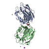
| Title | Vibrio cholerae GbpA (LPMO domain) | |||||||||
 Components Components | GlcNAc-binding protein A | |||||||||
 Keywords Keywords | CELL ADHESION / Adhesin / LPMO / redox enzyme / colonization factor | |||||||||
| Function / homology |  Function and homology information Function and homology information | |||||||||
| Biological species |  Vibrio cholerae O1 biovar El Tor (bacteria) Vibrio cholerae O1 biovar El Tor (bacteria) | |||||||||
| Method |  X-RAY DIFFRACTION / SYNCHROTRON / MOLECULAR REPLACEMENT / Resolution: 1.62 Å X-RAY DIFFRACTION / SYNCHROTRON / MOLECULAR REPLACEMENT / Resolution: 1.62 Å | |||||||||
 Authors Authors | Montserrat-Canals, M. / Sorensen, H.V. / Cordara, G. / Krengel, U. | |||||||||
| Funding support |  Norway, 2items Norway, 2items
| |||||||||
 Citation Citation | Journal: Acs Omega / Year: 2023 Title: Perdeuterated GbpA Enables Neutron Scattering Experiments of a Lytic Polysaccharide Monooxygenase. Authors: Sorensen, H.V. / Montserrat-Canals, M. / Loose, J.S.M. / Fisher, S.Z. / Moulin, M. / Blakeley, M.P. / Cordara, G. / Bjerregaard-Andersen, K. / Krengel, U. #1: Journal: Biochemistry / Year: 2023Title: Human 2'-Deoxynucleoside 5'-Phosphate N -Hydrolase 1: Mechanism of 2'-Deoxyuridine 5'-Monophosphate Hydrolysis. Authors: Devi, S. / Carberry, A.E. / Zickuhr, G.M. / Dickson, A.L. / Harrison, D.J. / da Silva, R.G. #2: Journal: Acs Omega / Year: 2023Title: Perdeuterated GbpA Enables Neutron Scattering Experiments of a Lytic Polysaccharide Monooxygenase. Authors: Sorensen, H.V. / Montserrat-Canals, M. / Loose, J.S.M. / Fisher, S.Z. / Moulin, M. / Blakeley, M.P. / Cordara, G. / Bjerregaard-Andersen, K. / Krengel, U. | |||||||||
| History |
|
- Structure visualization
| Structure viewer | Molecule: MolmilJmol/JSmol |
|---|
- Downloads & links
-Download
| PDBx/mmCIF format | 8cc5.cif.gz | 98.9 KB | Display | PDBx/mmCIF format |
|---|---|---|---|---|
| PDB format | pdb8cc5.ent.gz | 71.5 KB | Display | PDB format |
| PDBx/mmJSON format | 8cc5.json.gz | Tree view | PDBx/mmJSON format | |
| Others |  Other downloads Other downloads |
-Validation report
| Arichive directory | https://data.pdbj.org/pub/pdb/validation_reports/cc/8cc5ftp://data.pdbj.org/pub/pdb/validation_reports/cc/8cc5 | HTTPS FTP |
|---|
-Related structure data
-Links
 PDBj
PDBj- Assembly
| Deposited unit | 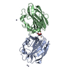
| ||||||||||||
|---|---|---|---|---|---|---|---|---|---|---|---|---|---|
| 1 | 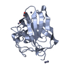
| ||||||||||||
| 2 | 
| ||||||||||||
| Unit cell |
| ||||||||||||
| Components on special symmetry positions |
|
-Components
-Protein , 1 types, 2 molecules AB
| #1: Protein | Mass: 19820.988 Da / Num. of mol.: 2 Source method: isolated from a genetically manipulated source Source: (gene. exp.) Vibrio cholerae O1 biovar El Tor (bacteria)Gene: gbpA, VCA0811 / Production host: |
|---|
-Non-polymers , 5 types, 200 molecules 


| #2: Chemical |  Mass: 59.044 Da / Num. of mol.: 2 / Source method: obtained synthetically / Formula: C2H3O2 Mass: 59.044 Da / Num. of mol.: 2 / Source method: obtained synthetically / Formula: C2H3O2#3: Chemical |  Mass: 63.546 Da / Num. of mol.: 2 / Source method: obtained synthetically / Formula: Cu / Feature type: SUBJECT OF INVESTIGATION Mass: 63.546 Da / Num. of mol.: 2 / Source method: obtained synthetically / Formula: Cu / Feature type: SUBJECT OF INVESTIGATION#4: Chemical | ChemComp-ZN /  Mass: 65.409 Da / Num. of mol.: 8 / Source method: obtained synthetically / Formula: Zn Mass: 65.409 Da / Num. of mol.: 8 / Source method: obtained synthetically / Formula: Zn#5: Chemical | ChemComp-NA / |  Mass: 22.990 Da / Num. of mol.: 1 / Source method: obtained synthetically / Formula: Na Mass: 22.990 Da / Num. of mol.: 1 / Source method: obtained synthetically / Formula: Na#6: Water | ChemComp-HOH / | Mass: 18.015 Da / Num. of mol.: 187 / Source method: isolated from a natural source / Formula: H2O |
|---|
-Details
| Has ligand of interest | Y |
|---|---|
| Has protein modification | Y |
-Experimental details
-Experiment
| Experiment | Method: X-RAY DIFFRACTION / Number of used crystals: 1 |
|---|
- Sample preparation
| Crystal | Density Matthews: 2.02 Å3/Da / Density % sol: 39.01 % |
|---|---|
| Crystal grow | Temperature: 293 K / Method: vapor diffusion, sitting drop / pH: 6.5 Details: 0.2 M Zinc Acetate 0.1 M Sodium Cacodylate pH 6.5 18% w/v PEG 8K |
-Data collection
| Diffraction | Mean temperature: 100 K / Serial crystal experiment: N |
|---|---|
| Diffraction source | Source: SYNCHROTRON / Site: ESRF  / Beamline: ID23-1 / Wavelength: 0.87313 Å / Beamline: ID23-1 / Wavelength: 0.87313 Å |
| Detector | Type: DECTRIS PILATUS3 2M / Detector: PIXEL / Date: Nov 29, 2021 |
| Radiation | Protocol: SINGLE WAVELENGTH / Monochromatic (M) / Laue (L): M / Scattering type: x-ray |
| Radiation wavelength | Wavelength: 0.87313 Å / Relative weight: 1 |
| Reflection | Resolution: 1.62→47.542 Å / Num. obs: 78744 / % possible obs: 99.94 % / Redundancy: 6.8 % / CC1/2: 0.998 / Net I/σ(I): 10.83 |
| Reflection shell | Resolution: 1.62→1.68 Å / Num. unique obs: 4085 / CC1/2: 0.374 |
- Processing
| Software |
| |||||||||||||||||||||||||||||||||||||||||||||||||||||||||||||||||||||||||||||||||||||||||||||||||||||||||||||||||||||||||||||||||||||||||||||||||||||||||||||||||||||||||||||||||||||||||||||
|---|---|---|---|---|---|---|---|---|---|---|---|---|---|---|---|---|---|---|---|---|---|---|---|---|---|---|---|---|---|---|---|---|---|---|---|---|---|---|---|---|---|---|---|---|---|---|---|---|---|---|---|---|---|---|---|---|---|---|---|---|---|---|---|---|---|---|---|---|---|---|---|---|---|---|---|---|---|---|---|---|---|---|---|---|---|---|---|---|---|---|---|---|---|---|---|---|---|---|---|---|---|---|---|---|---|---|---|---|---|---|---|---|---|---|---|---|---|---|---|---|---|---|---|---|---|---|---|---|---|---|---|---|---|---|---|---|---|---|---|---|---|---|---|---|---|---|---|---|---|---|---|---|---|---|---|---|---|---|---|---|---|---|---|---|---|---|---|---|---|---|---|---|---|---|---|---|---|---|---|---|---|---|---|---|---|---|---|---|---|---|
| Refinement | Method to determine structure: MOLECULAR REPLACEMENT / Resolution: 1.62→47.542 Å / Cor.coef. Fo:Fc: 0.97 / Cor.coef. Fo:Fc free: 0.959 / WRfactor Rfree: 0.208 / WRfactor Rwork: 0.172 / SU B: 2.11 / SU ML: 0.07 / Average fsc free: 0 / Average fsc work: 0 / Cross valid method: FREE R-VALUE / ESU R: 0.101 / ESU R Free: 0.101 Details: Hydrogens have been added in their riding positions
| |||||||||||||||||||||||||||||||||||||||||||||||||||||||||||||||||||||||||||||||||||||||||||||||||||||||||||||||||||||||||||||||||||||||||||||||||||||||||||||||||||||||||||||||||||||||||||||
| Solvent computation | Ion probe radii: 0.8 Å / Shrinkage radii: 0.8 Å / VDW probe radii: 1.2 Å / Solvent model: MASK BULK SOLVENT | |||||||||||||||||||||||||||||||||||||||||||||||||||||||||||||||||||||||||||||||||||||||||||||||||||||||||||||||||||||||||||||||||||||||||||||||||||||||||||||||||||||||||||||||||||||||||||||
| Displacement parameters | Biso mean: 25.413 Å2
| |||||||||||||||||||||||||||||||||||||||||||||||||||||||||||||||||||||||||||||||||||||||||||||||||||||||||||||||||||||||||||||||||||||||||||||||||||||||||||||||||||||||||||||||||||||||||||||
| Refinement step | Cycle: LAST / Resolution: 1.62→47.542 Å
| |||||||||||||||||||||||||||||||||||||||||||||||||||||||||||||||||||||||||||||||||||||||||||||||||||||||||||||||||||||||||||||||||||||||||||||||||||||||||||||||||||||||||||||||||||||||||||||
| Refine LS restraints |
| |||||||||||||||||||||||||||||||||||||||||||||||||||||||||||||||||||||||||||||||||||||||||||||||||||||||||||||||||||||||||||||||||||||||||||||||||||||||||||||||||||||||||||||||||||||||||||||
| LS refinement shell | Refine-ID: X-RAY DIFFRACTION / Total num. of bins used: 20
|
