Movie
Movie Controller

[English] 日本語
 Yorodumi
- PDB-8cbn: structure of LEDGF/p75 PWWP domain bound to the H3K36 trimethylat... -
+ Open data

- Basic information
| Entry | Database: PDB / ID: 8cbn | ||||||||||||
|---|---|---|---|---|---|---|---|---|---|---|---|---|---|
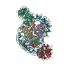
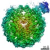
| Title | structure of LEDGF/p75 PWWP domain bound to the H3K36 trimethylated dinucleosome | ||||||||||||
 Components Components |
| ||||||||||||
 Keywords Keywords | TRANSCRIPTION / nucleosome / transcription activator / methylation / complex / DNA BINDING PROTEIN | ||||||||||||
| Function / homology |  Function and homology information Function and homology informationIntegration of viral DNA into host genomic DNA / Autointegration results in viral DNA circles / supercoiled DNA binding / 2-LTR circle formation / Vpr-mediated nuclear import of PICs / Formation of WDR5-containing histone-modifying complexes / mRNA 5'-splice site recognition / Integration of provirus / APOBEC3G mediated resistance to HIV-1 infection / heterochromatin ...Integration of viral DNA into host genomic DNA / Autointegration results in viral DNA circles / supercoiled DNA binding / 2-LTR circle formation / Vpr-mediated nuclear import of PICs / Formation of WDR5-containing histone-modifying complexes / mRNA 5'-splice site recognition / Integration of provirus / APOBEC3G mediated resistance to HIV-1 infection / heterochromatin / nuclear periphery / euchromatin / structural constituent of chromatin / nucleosome / nucleosome assembly / heterochromatin formation / response to heat / response to oxidative stress / DNA-binding transcription factor binding / transcription coactivator activity / chromatin remodeling / protein heterodimerization activity / chromatin binding / positive regulation of transcription by RNA polymerase II / DNA binding / RNA binding / nucleoplasm / nucleus / cytosol Similarity search - Function | ||||||||||||
| Biological species |  Homo sapiens (human) Homo sapiens (human)synthetic construct (others) | ||||||||||||
| Method | ELECTRON MICROSCOPY / single particle reconstruction / cryo EM / Resolution: 3.34 Å | ||||||||||||
 Authors Authors | Koutna, E. / Kouba, T. / Novacek, J. / Veverka, V. | ||||||||||||
| Funding support |  Czech Republic, European Union, 3items Czech Republic, European Union, 3items
| ||||||||||||
 Citation Citation | Journal: Nucleic Acids Res / Year: 2023 Title: Multivalency of nucleosome recognition by LEDGF. Authors: Eliška Koutná / Vanda Lux / Tomáš Kouba / Jana Škerlová / Jiří Nováček / Pavel Srb / Rozálie Hexnerová / Hana Šváchová / Zdeněk Kukačka / Petr Novák / Milan Fábry / Simon ...Authors: Eliška Koutná / Vanda Lux / Tomáš Kouba / Jana Škerlová / Jiří Nováček / Pavel Srb / Rozálie Hexnerová / Hana Šváchová / Zdeněk Kukačka / Petr Novák / Milan Fábry / Simon Poepsel / Václav Veverka /  Abstract: Eukaryotic transcription is dependent on specific histone modifications. Their recognition by chromatin readers triggers complex processes relying on the coordinated association of transcription ...Eukaryotic transcription is dependent on specific histone modifications. Their recognition by chromatin readers triggers complex processes relying on the coordinated association of transcription regulatory factors. Although various modification states of a particular histone residue often lead to differential outcomes, it is not entirely clear how they are discriminated. Moreover, the contribution of intrinsically disordered regions outside of the specialized reader domains to nucleosome binding remains unexplored. Here, we report the structures of a PWWP domain from transcriptional coactivator LEDGF in complex with the H3K36 di- and trimethylated nucleosome, indicating that both methylation marks are recognized by PWWP in a highly conserved manner. We identify a unique secondary interaction site for the PWWP domain at the interface between the acidic patch and nucleosomal DNA that might contribute to an H3K36-methylation independent role of LEDGF. We reveal DNA interacting motifs in the intrinsically disordered region of LEDGF that discriminate between the intra- or extranucleosomal DNA but remain dynamic in the context of dinucleosomes. The interplay between the LEDGF H3K36-methylation reader and protein binding module mediated by multivalent interactions of the intrinsically disordered linker with chromatin might help direct the elongation machinery to the vicinity of RNA polymerase II, thereby facilitating productive elongation. | ||||||||||||
| History |
|
- Structure visualization
| Structure viewer | Molecule: MolmilJmol/JSmol |
|---|
- Downloads & links
-Download
| PDBx/mmCIF format | 8cbn.cif.gz | 338.7 KB | Display | PDBx/mmCIF format |
|---|---|---|---|---|
| PDB format | pdb8cbn.ent.gz | 249 KB | Display | PDB format |
| PDBx/mmJSON format | 8cbn.json.gz | Tree view | PDBx/mmJSON format | |
| Others |  Other downloads Other downloads |
-Validation report
| Arichive directory | https://data.pdbj.org/pub/pdb/validation_reports/cb/8cbnftp://data.pdbj.org/pub/pdb/validation_reports/cb/8cbn | HTTPS FTP |
|---|
-Related structure data
| Related structure data | 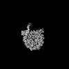 16546MC 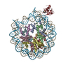 8cbqC 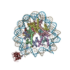 8pc5C 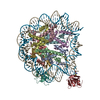 8pc6C 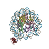 8peoC 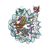 8pepC M: map data used to model this data C: citing same article ( |
|---|---|
| Similar structure data |


-Links
 PDBj
PDBj


- Assembly
| Deposited unit | 
|
|---|---|
| 1 |
|
-Components
-Protein , 5 types, 10 molecules BFCGDHKLAE
| #1: Protein | Mass: 11263.231 Da / Num. of mol.: 2 Source method: isolated from a genetically manipulated source Source: (gene. exp.)  #2: Protein | Mass: 13978.241 Da / Num. of mol.: 2 Source method: isolated from a genetically manipulated source Source: (gene. exp.) #3: Protein | Mass: 13524.752 Da / Num. of mol.: 2 Source method: isolated from a genetically manipulated source Source: (gene. exp.) #6: Protein | Mass: 60224.453 Da / Num. of mol.: 2 Source method: isolated from a genetically manipulated source Source: (gene. exp.) Homo sapiens (human) / Gene: PSIP1, DFS70, LEDGF, PSIP2 / Production host:  Trichoplusia ni (cabbage looper) / References: UniProt: O75475 Trichoplusia ni (cabbage looper) / References: UniProt: O75475#7: Protein | Mass: 15331.982 Da / Num. of mol.: 2 Source method: isolated from a genetically manipulated source Source: (gene. exp.) |
|---|
-DNA chain , 2 types, 2 molecules IJ
| #4: DNA chain | Mass: 50699.316 Da / Num. of mol.: 1 / Source method: obtained synthetically / Source: (synth.) synthetic construct (others) |
|---|---|
| #5: DNA chain | Mass: 51170.602 Da / Num. of mol.: 1 / Source method: obtained synthetically / Source: (synth.) synthetic construct (others) |
-Details
| Has ligand of interest | N |
|---|---|
| Has protein modification | Y |
-Experimental details
-Experiment
| Experiment | Method: ELECTRON MICROSCOPY |
|---|---|
| EM experiment | Aggregation state: PARTICLE / 3D reconstruction method: single particle reconstruction |
- Sample preparation
| Component |
| |||||||||||||||||||||||||||||||||||
|---|---|---|---|---|---|---|---|---|---|---|---|---|---|---|---|---|---|---|---|---|---|---|---|---|---|---|---|---|---|---|---|---|---|---|---|---|
| Molecular weight | Experimental value: NO | |||||||||||||||||||||||||||||||||||
| Source (natural) |
| |||||||||||||||||||||||||||||||||||
| Source (recombinant) |
| |||||||||||||||||||||||||||||||||||
| Buffer solution | pH: 7.5 | |||||||||||||||||||||||||||||||||||
| Specimen | Embedding applied: NO / Shadowing applied: NO / Staining applied: NO / Vitrification applied: YES | |||||||||||||||||||||||||||||||||||
| Vitrification | Cryogen name: ETHANE |
- Electron microscopy imaging
| Experimental equipment |  Model: Titan Krios / Image courtesy: FEI Company |
|---|---|
| Microscopy | Model: FEI TITAN KRIOS |
| Electron gun | Electron source:  FIELD EMISSION GUN / Accelerating voltage: 300 kV / Illumination mode: FLOOD BEAM FIELD EMISSION GUN / Accelerating voltage: 300 kV / Illumination mode: FLOOD BEAM |
| Electron lens | Mode: BRIGHT FIELD / Nominal defocus max: 3200 nm / Nominal defocus min: 1000 nm / Alignment procedure: COMA FREE |
| Specimen holder | Cryogen: NITROGEN |
| Image recording | Electron dose: 40 e/Å2 / Detector mode: COUNTING / Film or detector model: GATAN K2 SUMMIT (4k x 4k) |
- Processing
| EM software | Name: SerialEM / Category: image acquisition |
|---|---|
| CTF correction | Type: PHASE FLIPPING AND AMPLITUDE CORRECTION |
| 3D reconstruction | Resolution: 3.34 Å / Resolution method: FSC 0.143 CUT-OFF / Num. of particles: 154229 / Symmetry type: POINT |
| Atomic model building | Protocol: RIGID BODY FIT |
| Atomic model building | PDB-ID: 3MVD Accession code: 3MVD / Source name: PDB / Type: experimental model |
| Refinement | Highest resolution: 3.34 Å |