 Movie
Movie Controller

[English] 日本語
 Yorodumi
- PDB-8cb1: Crystal structure of human lysosomal acid-alpha-glucosidase, GAA,... -
+ Open data

- Basic information
| Entry | Database: PDB / ID: 8cb1 | ||||||
|---|---|---|---|---|---|---|---|
| Title | Crystal structure of human lysosomal acid-alpha-glucosidase, GAA, in complex with N-PNT-DNM 15 | ||||||
 Components Components | (Lysosomal alpha-glucosidase ...) x 2 | ||||||
 Keywords Keywords | HYDROLASE / Gaa / Pompe disease / pharmacological chaperone | ||||||
| Function / homology |  Function and homology information Function and homology information: / autolysosome lumen / maltose metabolic process / glucan 1,6-alpha-glucosidase activity / alpha-glucosidase activity / sucrose metabolic process / Glycogen storage disease type II (GAA) / alpha-glucosidase / alpha-1,4-glucosidase activity / neuromuscular process controlling posture ...: / autolysosome lumen / maltose metabolic process / glucan 1,6-alpha-glucosidase activity / alpha-glucosidase activity / sucrose metabolic process / Glycogen storage disease type II (GAA) / alpha-glucosidase / alpha-1,4-glucosidase activity / neuromuscular process controlling posture / glycophagy / diaphragm contraction / tissue development / regulation of the force of heart contraction / glycogen catabolic process / neuromuscular process controlling balance / aorta development / azurophil granule membrane / muscle cell cellular homeostasis / lysosome organization / Glycogen breakdown (glycogenolysis) / tertiary granule membrane / ficolin-1-rich granule membrane / heart morphogenesis / cardiac muscle contraction / lysosomal lumen / locomotory behavior / glucose metabolic process / carbohydrate binding / lysosome / lysosomal membrane / Neutrophil degranulation / extracellular exosome / membrane / plasma membrane Similarity search - Function | ||||||
| Biological species |  Homo sapiens (human) Homo sapiens (human) | ||||||
| Method |  X-RAY DIFFRACTION / SYNCHROTRON / FOURIER SYNTHESIS / Resolution: 1.75 Å X-RAY DIFFRACTION / SYNCHROTRON / FOURIER SYNTHESIS / Resolution: 1.75 Å | ||||||
 Authors Authors | Sulzenbacher, G. / Roig-Zamboni, V. / Overkleeft, H. / Artola, M. | ||||||
| Funding support | 1items
| ||||||
 Citation Citation | Journal: Chem Sci / Year: 2023 Title: Fluorescence polarisation activity-based protein profiling for the identification of deoxynojirimycin-type inhibitors selective for lysosomal retaining alpha- and beta-glucosidases. Authors: van der Gracht, D. / Rowland, R.J. / Roig-Zamboni, V. / Ferraz, M.J. / Louwerse, M. / Geurink, P.P. / Aerts, J.M.F.G. / Sulzenbacher, G. / Davies, G.J. / Overkleeft, H.S. / Artola, M. | ||||||
| History |
|
- Structure visualization
| Structure viewer | Molecule: MolmilJmol/JSmol |
|---|
- Downloads & links
-Download
| PDBx/mmCIF format | 8cb1.cif.gz | 218.1 KB | Display | PDBx/mmCIF format |
|---|---|---|---|---|
| PDB format | pdb8cb1.ent.gz | 164.6 KB | Display | PDB format |
| PDBx/mmJSON format | 8cb1.json.gz | Tree view | PDBx/mmJSON format | |
| Others |  Other downloads Other downloads |
-Validation report
| Arichive directory | https://data.pdbj.org/pub/pdb/validation_reports/cb/8cb1ftp://data.pdbj.org/pub/pdb/validation_reports/cb/8cb1 | HTTPS FTP |
|---|
-Related structure data
| Related structure data | 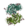 7nwvC  8cb6C  5nn3S S: Starting model for refinement C: citing same article ( |
|---|---|
| Similar structure data |
-Links
 PDBj
PDBj


- Assembly
| Deposited unit | 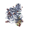
| ||||||||
|---|---|---|---|---|---|---|---|---|---|
| 1 |
| ||||||||
| Unit cell |
|
-Components
-Lysosomal alpha-glucosidase ... , 2 types, 2 molecules GA
| #1: Protein | Mass: 14095.020 Da / Num. of mol.: 1 Source method: isolated from a genetically manipulated source Details: FULL-LENGTH RHGAA HAS BEEN TREATED WITH CHYMOTRYPSIN, LEADING TO A SAMPLE STARTING AT RESIDUE GLN81. MISSING SURFACE LOOPS HAVE EQUALLY BEEN REMOVED BY PROTEOLYTIC CLEAVAGE. Source: (gene. exp.) Homo sapiens (human) / Gene: GAA / Production host:   Cricetulus griseus (Chinese hamster) / References: UniProt: P10253, alpha-glucosidase Cricetulus griseus (Chinese hamster) / References: UniProt: P10253, alpha-glucosidase |
|---|---|
| #2: Protein | Mass: 82901.570 Da / Num. of mol.: 1 Source method: isolated from a genetically manipulated source Details: FULL-LENGTH RHGAA HAS BEEN TREATED WITH CHYMOTRYPSIN, LEADING TO A SAMPLE STARTING AT RESIDUE GLN81. MISSING SURFACE LOOPS HAVE EQUALLY BEEN REMOVED BY PROTEOLYTIC CLEAVAGE. Source: (gene. exp.) Homo sapiens (human) / Gene: GAA / Production host: Cricetulus griseus (Chinese hamster) / References: UniProt: P10253 |
-Sugars , 4 types, 5 molecules
| #3: Polysaccharide | beta-D-mannopyranose-(1-4)-2-acetamido-2-deoxy-beta-D-glucopyranose-(1-4)-[alpha-L-fucopyranose-(1- ...beta-D-mannopyranose-(1-4)-2-acetamido-2-deoxy-beta-D-glucopyranose-(1-4)-[alpha-L-fucopyranose-(1-6)]2-acetamido-2-deoxy-beta-D-glucopyranose Source method: isolated from a genetically manipulated source | ||||
|---|---|---|---|---|---|
| #4: Polysaccharide | Source method: isolated from a genetically manipulated source #5: Polysaccharide | beta-D-mannopyranose-(1-4)-2-acetamido-2-deoxy-beta-D-glucopyranose-(1-4)-2-acetamido-2-deoxy-beta- ...beta-D-mannopyranose-(1-4)-2-acetamido-2-deoxy-beta-D-glucopyranose-(1-4)-2-acetamido-2-deoxy-beta-D-glucopyranose | Source method: isolated from a genetically manipulated source #6: Polysaccharide | 2-acetamido-2-deoxy-beta-D-glucopyranose-(1-4)-[alpha-L-fucopyranose-(1-6)]2-acetamido-2-deoxy-beta- ...2-acetamido-2-deoxy-beta-D-glucopyranose-(1-4)-[alpha-L-fucopyranose-(1-6)]2-acetamido-2-deoxy-beta-D-glucopyranose | Source method: isolated from a genetically manipulated source |
-Non-polymers , 7 types, 812 molecules 


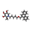


| #7: Chemical | ChemComp-CL /  Mass: 35.453 Da / Num. of mol.: 10 / Source method: obtained synthetically / Formula: Cl Mass: 35.453 Da / Num. of mol.: 10 / Source method: obtained synthetically / Formula: Cl#8: Chemical | ChemComp-GOL /  Mass: 92.094 Da / Num. of mol.: 4 / Source method: obtained synthetically / Formula: C3H8O3 Mass: 92.094 Da / Num. of mol.: 4 / Source method: obtained synthetically / Formula: C3H8O3#9: Chemical |  Mass: 150.173 Da / Num. of mol.: 3 / Source method: obtained synthetically / Formula: C6H14O4 / Feature type: SUBJECT OF INVESTIGATION Mass: 150.173 Da / Num. of mol.: 3 / Source method: obtained synthetically / Formula: C6H14O4 / Feature type: SUBJECT OF INVESTIGATION#10: Chemical | ChemComp-EDO /  Mass: 62.068 Da / Num. of mol.: 5 / Source method: obtained synthetically / Formula: C2H6O2 Mass: 62.068 Da / Num. of mol.: 5 / Source method: obtained synthetically / Formula: C2H6O2#11: Chemical | ChemComp-U4X / ( |  Mass: 439.544 Da / Num. of mol.: 1 / Source method: obtained synthetically / Formula: C26H33NO5 / Feature type: SUBJECT OF INVESTIGATION Mass: 439.544 Da / Num. of mol.: 1 / Source method: obtained synthetically / Formula: C26H33NO5 / Feature type: SUBJECT OF INVESTIGATION#12: Chemical | ChemComp-SO4 /  Mass: 96.063 Da / Num. of mol.: 4 / Source method: obtained synthetically / Formula: SO4 Mass: 96.063 Da / Num. of mol.: 4 / Source method: obtained synthetically / Formula: SO4#13: Water | ChemComp-HOH / | Mass: 18.015 Da / Num. of mol.: 785 / Source method: isolated from a natural source / Formula: H2O |
|---|
-Details
| Has ligand of interest | Y |
|---|---|
| Has protein modification | Y |
-Experimental details
-Experiment
| Experiment | Method: X-RAY DIFFRACTION / Number of used crystals: 1 |
|---|
- Sample preparation
| Crystal | Density Matthews: 3.4 Å3/Da / Density % sol: 62.79 % |
|---|---|
| Crystal grow | Temperature: 293 K / Method: vapor diffusion, hanging drop / pH: 7 Details: 1.9 M AMMONIUM SULPHATE, 0.1 M HEPES, 2% V/V PEG400 |
-Data collection
| Diffraction | Mean temperature: 100 K / Serial crystal experiment: N |
|---|---|
| Diffraction source | Source: SYNCHROTRON / Site: SOLEIL  / Beamline: PROXIMA 2 / Wavelength: 0.98011 Å / Beamline: PROXIMA 2 / Wavelength: 0.98011 Å |
| Detector | Type: DECTRIS EIGER X 9M / Detector: PIXEL / Date: Sep 15, 2021 |
| Radiation | Protocol: SINGLE WAVELENGTH / Monochromatic (M) / Laue (L): M / Scattering type: x-ray |
| Radiation wavelength | Wavelength: 0.98011 Å / Relative weight: 1 |
| Reflection | Resolution: 1.75→48.38 Å / Num. obs: 129900 / % possible obs: 100 % / Redundancy: 15.6 % / CC1/2: 0.999 / Rmerge(I) obs: 0.12 / Rpim(I) all: 0.031 / Rrim(I) all: 0.124 / Χ2: 0.97 / Net I/σ(I): 14.1 / Num. measured all: 2029400 |
| Reflection shell | Resolution: 1.75→1.78 Å / Redundancy: 15 % / Rmerge(I) obs: 2.065 / Num. unique obs: 6355 / CC1/2: 0.573 / Rpim(I) all: 0.537 / Rrim(I) all: 2.135 / Χ2: 0.91 |
- Processing
| Software |
| ||||||||||||||||||||||||||||||||||||||||||||||||||||||||||||||||||||||||||||||||||||||||||||||||||||||||||||||||||||||||||||||||||||||||||||||||||||||||||||||||||||||||||||||||||||||
|---|---|---|---|---|---|---|---|---|---|---|---|---|---|---|---|---|---|---|---|---|---|---|---|---|---|---|---|---|---|---|---|---|---|---|---|---|---|---|---|---|---|---|---|---|---|---|---|---|---|---|---|---|---|---|---|---|---|---|---|---|---|---|---|---|---|---|---|---|---|---|---|---|---|---|---|---|---|---|---|---|---|---|---|---|---|---|---|---|---|---|---|---|---|---|---|---|---|---|---|---|---|---|---|---|---|---|---|---|---|---|---|---|---|---|---|---|---|---|---|---|---|---|---|---|---|---|---|---|---|---|---|---|---|---|---|---|---|---|---|---|---|---|---|---|---|---|---|---|---|---|---|---|---|---|---|---|---|---|---|---|---|---|---|---|---|---|---|---|---|---|---|---|---|---|---|---|---|---|---|---|---|---|---|
| Refinement | Method to determine structure: FOURIER SYNTHESIS Starting model: 5NN3 Resolution: 1.75→47.73 Å / Cor.coef. Fo:Fc: 0.978 / Cor.coef. Fo:Fc free: 0.966 / SU B: 1.83 / SU ML: 0.056 / Cross valid method: THROUGHOUT / ESU R: 0.074 / ESU R Free: 0.079 / Stereochemistry target values: MAXIMUM LIKELIHOOD / Details: HYDROGENS HAVE BEEN ADDED IN THE RIDING POSITIONS
| ||||||||||||||||||||||||||||||||||||||||||||||||||||||||||||||||||||||||||||||||||||||||||||||||||||||||||||||||||||||||||||||||||||||||||||||||||||||||||||||||||||||||||||||||||||||
| Solvent computation | Ion probe radii: 0.8 Å / Shrinkage radii: 0.8 Å / VDW probe radii: 1.2 Å / Solvent model: BABINET MODEL WITH MASK | ||||||||||||||||||||||||||||||||||||||||||||||||||||||||||||||||||||||||||||||||||||||||||||||||||||||||||||||||||||||||||||||||||||||||||||||||||||||||||||||||||||||||||||||||||||||
| Displacement parameters | Biso mean: 40.587 Å2
| ||||||||||||||||||||||||||||||||||||||||||||||||||||||||||||||||||||||||||||||||||||||||||||||||||||||||||||||||||||||||||||||||||||||||||||||||||||||||||||||||||||||||||||||||||||||
| Refinement step | Cycle: 1 / Resolution: 1.75→47.73 Å
| ||||||||||||||||||||||||||||||||||||||||||||||||||||||||||||||||||||||||||||||||||||||||||||||||||||||||||||||||||||||||||||||||||||||||||||||||||||||||||||||||||||||||||||||||||||||
| Refine LS restraints |
|