[English] 日本語
Yorodumi
- PDB-8c64: Crystal structure of arsenoplatin-1/B-DNA adduct obtained upon 48... -

+
Open data


ID or keywords:

Loading...

-
Basic information

Entry
Database: PDB / ID: 8c64
TitleCrystal structure of arsenoplatin-1/B-DNA adduct obtained upon 48 h of soaking
ComponentsDNA (5'-D(*CP*GP*CP*GP*AP*AP*TP*TP*CP*GP*CP*G)-3')
KeywordsDNA / Duplex / B-DNA / Arsenoplatin-1 / Anticancer agent / Metallodrug
Function / homologyarsenoplatin-1 / : / DNA / DNA (> 10)
Function and homology information
Biological speciessynthetic construct (others)
MethodX-RAY DIFFRACTION / SYNCHROTRON / MOLECULAR REPLACEMENT / Resolution: 2.51 Å
AuthorsTroisi, R. / Tito, G. / Ferraro, G. / Sica, F. / Merlino, A.
Funding support1items
OrganizationGrant numberCountry
Not funded
CitationJournal: Dalton Trans / Year: 2024
Title: On the mechanism of action of arsenoplatins: arsenoplatin-1 binding to a B-DNA dodecamer.
Authors: Troisi, R. / Tito, G. / Ferraro, G. / Sica, F. / Massai, L. / Geri, A. / Cirri, D. / Messori, L. / Merlino, A.
History
DepositionJan 11, 2023Deposition site: PDBE / Processing site: PDBE
Revision 1.0Jan 31, 2024Provider: repository / Type: Initial release
Revision 1.1Feb 7, 2024Group: Database references / Category: citation / citation_author
Item: _citation.pdbx_database_id_DOI / _citation.pdbx_database_id_PubMed ..._citation.pdbx_database_id_DOI / _citation.pdbx_database_id_PubMed / _citation.title / _citation_author.identifier_ORCID
Revision 1.2Feb 28, 2024Group: Database references / Category: citation
Item: _citation.journal_volume / _citation.page_first / _citation.page_last

-
Structure visualization

Structure viewerMolecule:
MolmilJmol/JSmol

Downloads & links

-
Assembly

Deposited unit
A: DNA (5'-D(*CP*GP*CP*GP*AP*AP*TP*TP*CP*GP*CP*G)-3')
B: DNA (5'-D(*CP*GP*CP*GP*AP*AP*TP*TP*CP*GP*CP*G)-3')
hetero molecules


Theoretical massNumber of molelcules
Total (without water)8,1355
Polymers7,3272
Non-polymers8083
Water543
1


  • Idetical with deposited unit
  • defined by author&software
TypeNameSymmetry operationNumber
identity operation1_555x,y,z1
Buried area1480 Å2
ΔGint-48 kcal/mol
Surface area4500 Å2
MethodPISA
Unit cell
Length a, b, c (Å)39.970, 65.670, 23.560
Angle α, β, γ (deg.)90.00, 90.00, 90.00
Int Tables number19
Space group name H-MP212121

-
Components

#1: DNA chain DNA (5'-D(*CP*GP*CP*GP*AP*AP*TP*TP*CP*GP*CP*G)-3')


Mass: 3663.392 Da / Num. of mol.: 2 / Source method: obtained synthetically / Source: (synth.) synthetic construct (others)
#2: Chemical ChemComp-PT / PLATINUM (II) ION


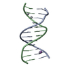
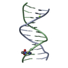
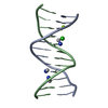
Mass: 195.078 Da / Num. of mol.: 2 / Source method: obtained synthetically / Formula: Pt / Feature type: SUBJECT OF INVESTIGATION
#3: Chemical ChemComp-A6R / arsenoplatin-1


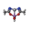
Mass: 418.117 Da / Num. of mol.: 1 / Source method: obtained synthetically / Formula: C4H8AsN2O4Pt / Feature type: SUBJECT OF INVESTIGATION
#4: Water ChemComp-HOH / water / Water


Mass: 18.015 Da / Num. of mol.: 3 / Source method: isolated from a natural source / Formula: H2O
Has ligand of interestY

-
Experimental details

-
Experiment

ExperimentMethod: X-RAY DIFFRACTION / Number of used crystals: 1

-
Sample preparation

CrystalDensity Matthews: 2.11 Å3/Da / Density % sol: 41.71 %
Crystal growTemperature: 293 K / Method: vapor diffusion, hanging drop / pH: 6.5
Details: MPD 7 % v/v, MgCl2 20 mM, spermine tetrahydrochloride 80 mM, sodium cacodylate 60 mM, pH 6.5. Equilibrated against a reservoir containing MPD 50 % v/v

-
Data collection

DiffractionMean temperature: 100 K / Serial crystal experiment: N
Diffraction sourceSource: SYNCHROTRON / Site: ELETTRA / Beamline: 11.2C / Wavelength: 1 Å
DetectorType: DECTRIS PILATUS 6M / Detector: PIXEL / Date: Jun 14, 2022
RadiationMonochromator: M / Protocol: SINGLE WAVELENGTH / Monochromatic (M) / Laue (L): M / Scattering type: x-ray
Radiation wavelengthWavelength: 1 Å / Relative weight: 1
ReflectionResolution: 2.51→34.14 Å / Num. obs: 2357 / % possible obs: 99.8 % / Redundancy: 10.5 % / CC1/2: 1 / Net I/σ(I): 13.7
Reflection shellResolution: 2.51→2.55 Å / Redundancy: 10.6 % / Mean I/σ(I) obs: 2.1 / Num. unique obs: 118 / CC1/2: 0.9 / % possible all: 100

-
Processing

Software
NameVersionClassification
REFMAC5.8.0403refinement
XDSdata reduction
XSCALEdata scaling
MOLREPphasing
autoPROCdata processing
RefinementMethod to determine structure: MOLECULAR REPLACEMENT / Resolution: 2.51→34.14 Å / Cor.coef. Fo:Fc: 0.975 / Cor.coef. Fo:Fc free: 0.963 / SU B: 8.944 / SU ML: 0.196 / Cross valid method: THROUGHOUT / ESU R: 1.085 / ESU R Free: 0.298 / Stereochemistry target values: MAXIMUM LIKELIHOOD
RfactorNum. reflection% reflectionSelection details
Rfree0.23238 91 4.2 %RANDOM
Rwork0.17865 ---
obs0.1807 2064 91.47 %-
Solvent computationIon probe radii: 0.8 Å / Shrinkage radii: 0.8 Å / VDW probe radii: 1.2 Å / Solvent model: MASK
Displacement parametersBiso mean: 59.082 Å2
Baniso -1Baniso -2Baniso -3
1-1.16 Å20 Å20 Å2
2---7.84 Å20 Å2
3---6.68 Å2
Refinement stepCycle: 1 / Resolution: 2.51→34.14 Å
ProteinNucleic acidLigandSolventTotal
Num. atoms0 486 4 3 493
Refine LS restraints
Refine-IDTypeDev idealDev ideal targetNumber
X-RAY DIFFRACTIONr_bond_refined_d0.0070.011545
X-RAY DIFFRACTIONr_bond_other_d
X-RAY DIFFRACTIONr_angle_refined_deg2.3441.786836
X-RAY DIFFRACTIONr_angle_other_deg
X-RAY DIFFRACTIONr_dihedral_angle_1_deg
X-RAY DIFFRACTIONr_dihedral_angle_2_deg
X-RAY DIFFRACTIONr_dihedral_angle_3_deg
X-RAY DIFFRACTIONr_dihedral_angle_4_deg
X-RAY DIFFRACTIONr_chiral_restr0.0840.294
X-RAY DIFFRACTIONr_gen_planes_refined0.0150.02252
X-RAY DIFFRACTIONr_gen_planes_other
X-RAY DIFFRACTIONr_nbd_refined
X-RAY DIFFRACTIONr_nbd_other
X-RAY DIFFRACTIONr_nbtor_refined
X-RAY DIFFRACTIONr_nbtor_other
X-RAY DIFFRACTIONr_xyhbond_nbd_refined
X-RAY DIFFRACTIONr_xyhbond_nbd_other
X-RAY DIFFRACTIONr_metal_ion_refined
X-RAY DIFFRACTIONr_metal_ion_other
X-RAY DIFFRACTIONr_symmetry_vdw_refined
X-RAY DIFFRACTIONr_symmetry_vdw_other
X-RAY DIFFRACTIONr_symmetry_hbond_refined
X-RAY DIFFRACTIONr_symmetry_hbond_other
X-RAY DIFFRACTIONr_symmetry_metal_ion_refined
X-RAY DIFFRACTIONr_symmetry_metal_ion_other
X-RAY DIFFRACTIONr_mcbond_it
X-RAY DIFFRACTIONr_mcbond_other
X-RAY DIFFRACTIONr_mcangle_it
X-RAY DIFFRACTIONr_mcangle_other
X-RAY DIFFRACTIONr_scbond_it7.7046.058545
X-RAY DIFFRACTIONr_scbond_other
X-RAY DIFFRACTIONr_scangle_it
X-RAY DIFFRACTIONr_scangle_other
X-RAY DIFFRACTIONr_long_range_B_refined12.35586.69966
X-RAY DIFFRACTIONr_long_range_B_other
X-RAY DIFFRACTIONr_rigid_bond_restr
X-RAY DIFFRACTIONr_sphericity_free
X-RAY DIFFRACTIONr_sphericity_bonded
LS refinement shellResolution: 2.511→2.576 Å / Total num. of bins used: 20
RfactorNum. reflection% reflection
Rfree0.392 8 -
Rwork0.293 155 -
obs--92.09 %

+
About Yorodumi

-
News

-
Feb 9, 2022. New format data for meta-information of EMDB entries

New format data for meta-information of EMDB entries

  • Version 3 of the EMDB header file is now the official format.
  • The previous official version 1.9 will be removed from the archive.

Related info.:EMDB header

External links:wwPDB to switch to version 3 of the EMDB data model

-
Aug 12, 2020. Covid-19 info

Covid-19 info

URL: https://pdbj.org/emnavi/covid19.php

New page: Covid-19 featured information page in EM Navigator.

Related info.:Covid-19 info / Mar 5, 2020. Novel coronavirus structure data

+
Mar 5, 2020. Novel coronavirus structure data

Novel coronavirus structure data

Related info.:Yorodumi Speices / Aug 12, 2020. Covid-19 info

External links:COVID-19 featured content - PDBj / Molecule of the Month (242):Coronavirus Proteases

+
Jan 31, 2019. EMDB accession codes are about to change! (news from PDBe EMDB page)

EMDB accession codes are about to change! (news from PDBe EMDB page)

  • The allocation of 4 digits for EMDB accession codes will soon come to an end. Whilst these codes will remain in use, new EMDB accession codes will include an additional digit and will expand incrementally as the available range of codes is exhausted. The current 4-digit format prefixed with “EMD-” (i.e. EMD-XXXX) will advance to a 5-digit format (i.e. EMD-XXXXX), and so on. It is currently estimated that the 4-digit codes will be depleted around Spring 2019, at which point the 5-digit format will come into force.
  • The EM Navigator/Yorodumi systems omit the EMD- prefix.

Related info.:Q: What is EMD? / ID/Accession-code notation in Yorodumi/EM Navigator

External links:EMDB Accession Codes are Changing Soon! / Contact to PDBj

+
Jul 12, 2017. Major update of PDB

Major update of PDB

  • wwPDB released updated PDB data conforming to the new PDBx/mmCIF dictionary.
  • This is a major update changing the version number from 4 to 5, and with Remediation, in which all the entries are updated.
  • In this update, many items about electron microscopy experimental information are reorganized (e.g. em_software).
  • Now, EM Navigator and Yorodumi are based on the updated data.

External links:wwPDB Remediation / Enriched Model Files Conforming to OneDep Data Standards Now Available in the PDB FTP Archive

-
Yorodumi

Thousand views of thousand structures

  • Yorodumi is a browser for structure data from EMDB, PDB, SASBDB, etc.
  • This page is also the successor to EM Navigator detail page, and also detail information page/front-end page for Omokage search.
  • The word "yorodu" (or yorozu) is an old Japanese word meaning "ten thousand". "mi" (miru) is to see.

Related info.:EMDB / PDB / SASBDB / Comparison of 3 databanks / Yorodumi Search / Aug 31, 2016. New EM Navigator & Yorodumi / Yorodumi Papers / Jmol/JSmol / Function and homology information / Changes in new EM Navigator and Yorodumi

Read more