 Movie
Movie Controller

[English] 日本語
 Yorodumi
- PDB-8c5i: Cyanide dihydratase from Bacillus pumilus C1 variant - Q86R,H305K... -
+ Open data

- Basic information
| Entry | Database: PDB / ID: 8c5i | ||||||
|---|---|---|---|---|---|---|---|
| Title | Cyanide dihydratase from Bacillus pumilus C1 variant - Q86R,H305K,H308K,H323K | ||||||
 Components Components | Cyanide dihydratase | ||||||
 Keywords Keywords | HYDROLASE / Helical / homo-oligomeric / cyanide dihydratase | ||||||
| Function / homology |  Function and homology information Function and homology informationnitrilase activity / detoxification of nitrogen compound / nitrile hydratase activity Similarity search - Function | ||||||
| Biological species |  | ||||||
| Method | ELECTRON MICROSCOPY / helical reconstruction / cryo EM / Resolution: 3.15 Å | ||||||
 Authors Authors | Mulelu, A.E. / Reitz, J. / van Rooyen, J. / Scheffer, M. / Frangakis, A.S. / Dlamini, L.S. / Woodward, J.D. / Benedik, M.J. / Sewell, B.T. | ||||||
| Funding support |  South Africa, 1items South Africa, 1items
| ||||||
 Citation Citation | Journal: To Be Published Title: The Role of Histidine Residues in the Oligomerization of Cyanide Dihydratase from Bacillus pumilus C1 Authors: Mulelu, A.E. / Reitz, J. / van Rooyen, J. / Scheffer, M. / Frangakis, A.S. / Dlamini, L.S. / Woodward, J.D. / Benedik, M.J. / Sewell, B.T. | ||||||
| History |
|
- Structure visualization
| Structure viewer | Molecule: MolmilJmol/JSmol |
|---|
- Downloads & links
-Download
| PDBx/mmCIF format | 8c5i.cif.gz | 2 MB | Display | PDBx/mmCIF format |
|---|---|---|---|---|
| PDB format | pdb8c5i.ent.gz | 1.7 MB | Display | PDB format |
| PDBx/mmJSON format | 8c5i.json.gz | Tree view | PDBx/mmJSON format | |
| Others |  Other downloads Other downloads |
-Validation report
| Arichive directory | https://data.pdbj.org/pub/pdb/validation_reports/c5/8c5iftp://data.pdbj.org/pub/pdb/validation_reports/c5/8c5i | HTTPS FTP |
|---|
-Related structure data
| Related structure data | 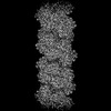 16437MC 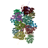 8p4iC M: map data used to model this data C: citing same article ( |
|---|---|
| Similar structure data |
-Links
 PDBj
PDBj- Assembly
| Deposited unit | 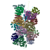
|
|---|---|
| 1 |
|
-Components
| #1: Protein | Mass: 37503.363 Da / Num. of mol.: 18 / Mutation: Q86R,H305K,H308K,H323K Source method: isolated from a genetically manipulated source Source: (gene. exp.) |
|---|
-Experimental details
-Experiment
| Experiment | Method: ELECTRON MICROSCOPY |
|---|---|
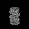
| EM experiment | Aggregation state: FILAMENT / 3D reconstruction method: helical reconstruction |
- Sample preparation
| Component | Name: Active helical nitrilase homo-oligomer of cyanide dihydratase from Bacillus pumilus C1 variant (Q86R/H305K/H308K/H323K) Type: COMPLEX Details: Cyanide dihydratase from Bacillus pumilus C1 variant generated by site-directed mutagenesis. Entity ID: all / Source: RECOMBINANT | |||||||||||||||
|---|---|---|---|---|---|---|---|---|---|---|---|---|---|---|---|---|
| Molecular weight | Experimental value: NO | |||||||||||||||
| Source (natural) | Organism: | |||||||||||||||
| Source (recombinant) | Organism: | |||||||||||||||
| Buffer solution | pH: 5.4 / Details: 150 mM NaCl, 50 mM Tris-HCl pH 5.4 | |||||||||||||||
| Buffer component |
| |||||||||||||||
| Specimen | Conc.: 0.2 mg/ml / Embedding applied: NO / Shadowing applied: NO / Staining applied: NO / Vitrification applied: YES / Details: Homogeneous protein sample. | |||||||||||||||
| Specimen support | Grid material: COPPER / Grid type: Quantifoil R2/2 | |||||||||||||||
| Vitrification | Instrument: FEI VITROBOT MARK IV / Cryogen name: ETHANE / Humidity: 100 % / Chamber temperature: 277.15 K Details: A 2.5 microlitre sample was applied onto a glow-discharged grid, blotted and plunged without incubation. |
- Electron microscopy imaging
| Experimental equipment |  Model: Titan Krios / Image courtesy: FEI Company |
|---|---|
| Microscopy | Model: FEI TITAN KRIOS |
| Electron gun | Electron source:  FIELD EMISSION GUN / Accelerating voltage: 300 kV / Illumination mode: OTHER FIELD EMISSION GUN / Accelerating voltage: 300 kV / Illumination mode: OTHER |
| Electron lens | Mode: BRIGHT FIELD / Nominal magnification: 130000 X / Nominal defocus max: 3000 nm / Nominal defocus min: 600 nm |
| Specimen holder | Cryogen: NITROGEN / Specimen holder model: FEI TITAN KRIOS AUTOGRID HOLDER |
| Image recording | Electron dose: 54 e/Å2 / Detector mode: COUNTING / Film or detector model: GATAN K2 SUMMIT (4k x 4k) |
| Image scans | Movie frames/image: 30 |
- Processing
| Software | Name: PHENIX / Version: 1.20.1_4487: / Classification: refinement | |||||||||||||||||||||||||||||||||||
|---|---|---|---|---|---|---|---|---|---|---|---|---|---|---|---|---|---|---|---|---|---|---|---|---|---|---|---|---|---|---|---|---|---|---|---|---|
| EM software |
| |||||||||||||||||||||||||||||||||||
| CTF correction | Type: PHASE FLIPPING ONLY | |||||||||||||||||||||||||||||||||||
| Helical symmerty | Angular rotation/subunit: -77 ° / Axial rise/subunit: 16.7 Å / Axial symmetry: C2 | |||||||||||||||||||||||||||||||||||
| 3D reconstruction | Resolution: 3.15 Å / Resolution method: FSC 0.5 CUT-OFF / Num. of particles: 103000 / Algorithm: FOURIER SPACE / Num. of class averages: 92000 / Symmetry type: HELICAL | |||||||||||||||||||||||||||||||||||
| Atomic model building | Protocol: AB INITIO MODEL / Space: REAL | |||||||||||||||||||||||||||||||||||
| Refine LS restraints |
|
