 Movie
Movie Controller

[English] 日本語
 Yorodumi
- PDB-8c5c: microtubule decorated with tubulin oligomers in presence of APC C... -
+ Open data

- Basic information
| Entry | Database: PDB / ID: 8c5c | |||||||||||||||||||||||||||||||||||||||
|---|---|---|---|---|---|---|---|---|---|---|---|---|---|---|---|---|---|---|---|---|---|---|---|---|---|---|---|---|---|---|---|---|---|---|---|---|---|---|---|---|
| Title | microtubule decorated with tubulin oligomers in presence of APC C-terminal domain. (here only map corresponding to the 13-pf microtubule is represented) | |||||||||||||||||||||||||||||||||||||||
 Components Components |
| |||||||||||||||||||||||||||||||||||||||
 Keywords Keywords | CELL CYCLE / cytoskeleton / microtubule | |||||||||||||||||||||||||||||||||||||||
| Function / homology |  Function and homology information Function and homology informationcytoplasmic microtubule / cellular response to interleukin-4 / structural constituent of cytoskeleton / microtubule cytoskeleton organization / neuron migration / mitotic cell cycle / double-stranded RNA binding / microtubule cytoskeleton / microtubule / Hydrolases; Acting on acid anhydrides; Acting on GTP to facilitate cellular and subcellular movement ...cytoplasmic microtubule / cellular response to interleukin-4 / structural constituent of cytoskeleton / microtubule cytoskeleton organization / neuron migration / mitotic cell cycle / double-stranded RNA binding / microtubule cytoskeleton / microtubule / Hydrolases; Acting on acid anhydrides; Acting on GTP to facilitate cellular and subcellular movement / cilium / GTPase activity / ubiquitin protein ligase binding / GTP binding / metal ion binding / cytoplasm / cytosol Similarity search - Function | |||||||||||||||||||||||||||||||||||||||
| Biological species |  | |||||||||||||||||||||||||||||||||||||||
| Method | ELECTRON MICROSCOPY / helical reconstruction / cryo EM / Resolution: 5.3 Å | |||||||||||||||||||||||||||||||||||||||
 Authors Authors | Serre, L. / Arnal, I. | |||||||||||||||||||||||||||||||||||||||
| Funding support |  France, 1items France, 1items
| |||||||||||||||||||||||||||||||||||||||
 Citation Citation | Journal: J Cell Sci / Year: 2023 Title: The mitotic role of adenomatous polyposis coli requires its bilateral interaction with tubulin and microtubules. Authors: Laurence Serre / Julie Delaroche / Angélique Vinit / Guy Schoehn / Eric Denarier / Anne Fourest-Lieuvin / Isabelle Arnal / Abstract: Adenomatous polyposis coli (APC) is a scaffold protein with tumour suppressor properties. Mutations causing the loss of its C-terminal domain (APC-C), which bears cytoskeleton-regulating sequences, ...Adenomatous polyposis coli (APC) is a scaffold protein with tumour suppressor properties. Mutations causing the loss of its C-terminal domain (APC-C), which bears cytoskeleton-regulating sequences, correlate with colorectal cancer. The cellular roles of APC in mitosis are widely studied, but the molecular mechanisms of its interaction with the cytoskeleton are poorly understood. Here, we investigated how APC-C regulates microtubule properties, and found that it promotes both microtubule growth and shrinkage. Strikingly, APC-C accumulates at shrinking microtubule extremities, a common characteristic of depolymerases. Cryo-electron microscopy revealed that APC-C adopts an extended conformation along the protofilament crest and showed the presence of ring-like tubulin oligomers around the microtubule wall, which required the presence of two APC-C sub-domains. A mutant of APC-C that was incapable of decorating microtubules with ring-like tubulin oligomers exhibited a reduced effect on microtubule dynamics. Finally, whereas native APC-C rescued defective chromosome alignment in metaphase cells silenced for APC, the ring-incompetent mutant failed to correct mitotic defects. Thus, the bilateral interaction of APC-C with tubulin and microtubules likely contributes to its mitotic functions. | |||||||||||||||||||||||||||||||||||||||
| History |
|
- Structure visualization
| Structure viewer | Molecule: MolmilJmol/JSmol |
|---|
- Downloads & links
-Download
| PDBx/mmCIF format | 8c5c.cif.gz | 4.2 MB | Display | PDBx/mmCIF format |
|---|---|---|---|---|
| PDB format | pdb8c5c.ent.gz | Display | PDB format | |
| PDBx/mmJSON format | 8c5c.json.gz | Tree view | PDBx/mmJSON format | |
| Others |  Other downloads Other downloads |
-Validation report
| Arichive directory | https://data.pdbj.org/pub/pdb/validation_reports/c5/8c5cftp://data.pdbj.org/pub/pdb/validation_reports/c5/8c5c | HTTPS FTP |
|---|
-Related structure data
| Related structure data | 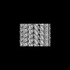 16435MC M: map data used to model this data C: citing same article ( |
|---|---|
| Similar structure data |

-Links
 PDBj
PDBj


- Assembly
| Deposited unit | 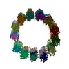
|
|---|---|
| 1 |
|
-Components
-Protein , 2 types, 52 molecules abcdefghijklmnopqrstuvwxyzNOPQ...
| #1: Protein | Mass: 50019.922 Da / Num. of mol.: 26 / Source method: isolated from a natural source / Source: (natural) #2: Protein | Mass: 50204.445 Da / Num. of mol.: 26 / Source method: isolated from a natural source / Source: (natural) |
|---|
-Non-polymers , 4 types, 52 molecules 


| #3: Chemical | ChemComp-TA1 /  Mass: 853.906 Da / Num. of mol.: 13 / Source method: obtained synthetically / Formula: C47H51NO14 / Feature type: SUBJECT OF INVESTIGATION / Comment: medication*YM Mass: 853.906 Da / Num. of mol.: 13 / Source method: obtained synthetically / Formula: C47H51NO14 / Feature type: SUBJECT OF INVESTIGATION / Comment: medication*YM#4: Chemical | ChemComp-GDP /  Type: RNA linking / Mass: 443.201 Da / Num. of mol.: 13 / Source method: obtained synthetically / Formula: C10H15N5O11P2 / Feature type: SUBJECT OF INVESTIGATION / Comment: GDP, energy-carrying molecule*YM Type: RNA linking / Mass: 443.201 Da / Num. of mol.: 13 / Source method: obtained synthetically / Formula: C10H15N5O11P2 / Feature type: SUBJECT OF INVESTIGATION / Comment: GDP, energy-carrying molecule*YM#5: Chemical | ChemComp-MG /  Mass: 24.305 Da / Num. of mol.: 13 / Source method: obtained synthetically / Formula: Mg / Feature type: SUBJECT OF INVESTIGATION Mass: 24.305 Da / Num. of mol.: 13 / Source method: obtained synthetically / Formula: Mg / Feature type: SUBJECT OF INVESTIGATION#6: Chemical | ChemComp-GTP /  Mass: 523.180 Da / Num. of mol.: 13 / Source method: obtained synthetically / Formula: C10H16N5O14P3 / Feature type: SUBJECT OF INVESTIGATION / Comment: GTP, energy-carrying molecule*YM Mass: 523.180 Da / Num. of mol.: 13 / Source method: obtained synthetically / Formula: C10H16N5O14P3 / Feature type: SUBJECT OF INVESTIGATION / Comment: GTP, energy-carrying molecule*YM |
|---|
-Details
| Has ligand of interest | Y |
|---|---|
| Has protein modification | N |
-Experimental details
-Experiment
| Experiment | Method: ELECTRON MICROSCOPY |
|---|---|
| EM experiment | Aggregation state: FILAMENT / 3D reconstruction method: helical reconstruction |
- Sample preparation
| Component | Name: microtubule / Type: COMPLEX / Entity ID: #1-#2 / Source: NATURAL | |||||||||||||||||||||||||
|---|---|---|---|---|---|---|---|---|---|---|---|---|---|---|---|---|---|---|---|---|---|---|---|---|---|---|
| Source (natural) | Organism: | |||||||||||||||||||||||||
| Buffer solution | pH: 6.75 | |||||||||||||||||||||||||
| Buffer component |
| |||||||||||||||||||||||||
| Specimen | Embedding applied: NO / Shadowing applied: NO / Staining applied: NO / Vitrification applied: YES | |||||||||||||||||||||||||
| Specimen support | Grid material: COPPER / Grid mesh size: 300 divisions/in. / Grid type: Quantifoil R2/2 | |||||||||||||||||||||||||
| Vitrification | Instrument: FEI VITROBOT MARK IV / Cryogen name: ETHANE / Humidity: 100 % / Chamber temperature: 37 K |
- Electron microscopy imaging
| Microscopy | Model: TFS GLACIOS |
|---|---|
| Electron gun | Electron source:  FIELD EMISSION GUN / Accelerating voltage: 200 kV / Illumination mode: FLOOD BEAM FIELD EMISSION GUN / Accelerating voltage: 200 kV / Illumination mode: FLOOD BEAM |
| Electron lens | Mode: BRIGHT FIELD / Nominal defocus max: 3000 nm / Nominal defocus min: 1500 nm |
| Image recording | Electron dose: 40 e/Å2 / Film or detector model: FEI FALCON II (4k x 4k) |
- Processing
| EM software |
| ||||||||||||
|---|---|---|---|---|---|---|---|---|---|---|---|---|---|
| CTF correction | Type: PHASE FLIPPING AND AMPLITUDE CORRECTION | ||||||||||||
| Helical symmerty | Angular rotation/subunit: -27.68 ° / Axial rise/subunit: 9.54 Å / Axial symmetry: C1 | ||||||||||||
| 3D reconstruction | Resolution: 5.3 Å / Resolution method: FSC 0.143 CUT-OFF / Num. of particles: 11670 / Details: resolution deduced from unmasked FSC is 7.1 A / Symmetry type: HELICAL |
