 Movie
Movie Controller

[English] 日本語
 Yorodumi
- PDB-8c53: Trypanosoma brucei IMP dehydrogenase (ori) crystallized in High F... -
+ Open data

- Basic information
| Entry | Database: PDB / ID: 8c53 | ||||||||||||
|---|---|---|---|---|---|---|---|---|---|---|---|---|---|
| Title | Trypanosoma brucei IMP dehydrogenase (ori) crystallized in High Five cells reveals native ligands ATP, GDP and phosphate. Diffraction data collection at 100 K in cellulo; CrystFEL processing | ||||||||||||
 Components Components | Inosine-5'-monophosphate dehydrogenase | ||||||||||||
 Keywords Keywords | TRANSFERASE / IMP dehydrogenase / purine biosynthetic enzyme / native cofactors | ||||||||||||
| Function / homology |  Function and homology information Function and homology informationIMP dehydrogenase / glycosome / IMP dehydrogenase activity / GMP biosynthetic process / GTP biosynthetic process / nucleotide binding / metal ion binding Similarity search - Function | ||||||||||||
| Biological species |  | ||||||||||||
| Method |  X-RAY DIFFRACTION / SYNCHROTRON / MOLECULAR REPLACEMENT / Resolution: 2.3 Å X-RAY DIFFRACTION / SYNCHROTRON / MOLECULAR REPLACEMENT / Resolution: 2.3 Å | ||||||||||||
 Authors Authors | Lahey-Rudolph, J.M. / Schoenherr, R. / Boger, J. / Harms, M. / Kaiser, J. / Nachtschatt, S. / Wobbe, M. / Duden, R. / Bourenkov, G. / Schneider, T. / Redecke, L. | ||||||||||||
| Funding support |  Germany, 3items Germany, 3items
| ||||||||||||
 Citation Citation | Journal: Nat Commun / Year: 2024 Title: A streamlined approach to structure elucidation using in cellulo crystallized recombinant proteins, InCellCryst. Authors: Schonherr, R. / Boger, J. / Lahey-Rudolph, J.M. / Harms, M. / Kaiser, J. / Nachtschatt, S. / Wobbe, M. / Duden, R. / Konig, P. / Bourenkov, G. / Schneider, T.R. / Redecke, L. | ||||||||||||
| History |
|
- Structure visualization
| Structure viewer | Molecule: MolmilJmol/JSmol |
|---|
- Downloads & links
-Download
| PDBx/mmCIF format | 8c53.cif.gz | 337.9 KB | Display | PDBx/mmCIF format |
|---|---|---|---|---|
| PDB format | pdb8c53.ent.gz | 276.5 KB | Display | PDB format |
| PDBx/mmJSON format | 8c53.json.gz | Tree view | PDBx/mmJSON format | |
| Others |  Other downloads Other downloads |
-Validation report
| Summary document | 8c53_validation.pdf.gz | 2.8 MB | Display | wwPDB validaton report |
|---|---|---|---|---|
| Full document | 8c53_full_validation.pdf.gz | 2.8 MB | Display | |
| Data in XML | 8c53_validation.xml.gz | 35.4 KB | Display | |
| Data in CIF | 8c53_validation.cif.gz | 48.2 KB | Display | |
| Arichive directory | https://data.pdbj.org/pub/pdb/validation_reports/c5/8c53ftp://data.pdbj.org/pub/pdb/validation_reports/c5/8c53 | HTTPS FTP |
-Related structure data
| Related structure data | 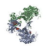 8c51C  8c5kC 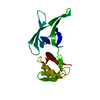 8cd4C 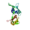 8cd5C 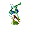 8cd6C  8cgxC 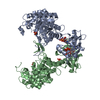 8cgyC  1jcnS C: citing same article ( S: Starting model for refinement |
|---|---|
| Similar structure data | |
| Other databases |
|
-Links
 PDBj
PDBj
- Assembly
| Deposited unit | 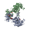
| ||||||||||
|---|---|---|---|---|---|---|---|---|---|---|---|
| 1 | 
| ||||||||||
| Unit cell |
| ||||||||||
| Noncrystallographic symmetry (NCS) | NCS oper: (Code: givenMatrix: (0.498633116137, -0.86677619989, -0.00800217442322), (-0.866793669248, -0.498662541492, 0.00209872922587), (-0.00580951317835, 0.00588973823644, -0.999965779685)Vector: ...NCS oper: (Code: given Matrix: (0.498633116137, -0.86677619989, -0.00800217442322), Vector: |
-Components
| #1: Protein | Mass: 55765.656 Da / Num. of mol.: 2 / Mutation: T488S Source method: isolated from a genetically manipulated source Source: (gene. exp.)  Trichoplusia ni (cabbage looper) / References: UniProt: P50098, IMP dehydrogenase Trichoplusia ni (cabbage looper) / References: UniProt: P50098, IMP dehydrogenase#2: Chemical |   Type: RNA linking / Mass: 443.201 Da / Num. of mol.: 2 / Source method: obtained synthetically / Formula: C10H15N5O11P2 / Feature type: SUBJECT OF INVESTIGATION / Comment: GDP, energy-carrying molecule*YM Type: RNA linking / Mass: 443.201 Da / Num. of mol.: 2 / Source method: obtained synthetically / Formula: C10H15N5O11P2 / Feature type: SUBJECT OF INVESTIGATION / Comment: GDP, energy-carrying molecule*YM#3: Chemical |   Mass: 507.181 Da / Num. of mol.: 2 / Source method: obtained synthetically / Formula: C10H16N5O13P3 / Feature type: SUBJECT OF INVESTIGATION / Comment: ATP, energy-carrying molecule*YM Mass: 507.181 Da / Num. of mol.: 2 / Source method: obtained synthetically / Formula: C10H16N5O13P3 / Feature type: SUBJECT OF INVESTIGATION / Comment: ATP, energy-carrying molecule*YM#4: Chemical |   Mass: 94.971 Da / Num. of mol.: 2 / Source method: isolated from a natural source / Formula: PO4 / Feature type: SUBJECT OF INVESTIGATION Mass: 94.971 Da / Num. of mol.: 2 / Source method: isolated from a natural source / Formula: PO4 / Feature type: SUBJECT OF INVESTIGATION#5: Water | ChemComp-HOH / |  Mass: 18.015 Da / Num. of mol.: 150 / Source method: isolated from a natural source / Formula: H2O Mass: 18.015 Da / Num. of mol.: 150 / Source method: isolated from a natural source / Formula: H2OHas ligand of interest | Y | |
|---|
-Experimental details
-Experiment
| Experiment | Method: X-RAY DIFFRACTION / Number of used crystals: 1 |
|---|
- Sample preparation
| Crystal | Density Matthews: 4.43 Å3/Da / Density % sol: 72.22 % |
|---|---|
| Crystal grow | Temperature: 300 K / Method: in cell Details: crystallized in rBV Tb IMPDH ori (MOI 1) infected High Five cells, in adhesion culture using ESF921 cell culture medium. |
-Data collection
| Diffraction | Mean temperature: 100 K / Serial crystal experiment: Y |
|---|---|
| Diffraction source | Source: SYNCHROTRON / Site: PETRA III, EMBL c/o DESY / Beamline: P14 (MX2) / Wavelength: 0.987 Å |
| Detector | Type: DECTRIS EIGER2 S 16M / Detector: PIXEL / Date: Nov 26, 2019 |
| Radiation | Protocol: SINGLE WAVELENGTH / Monochromatic (M) / Laue (L): M / Scattering type: x-ray |
| Radiation wavelength | Wavelength: 0.987 Å / Relative weight: 1 |
| Reflection | Resolution: 2.299→57.38 Å / Num. obs: 89070 / % possible obs: 99.88 % / Redundancy: 3667 % / Biso Wilson estimate: 52.99 Å2 / CC1/2: 0.9976 / CC star: 0.9994 / R split: 0.0862 / Net I/σ(I): 10.06 |
| Reflection shell | Resolution: 2.299→2.33 Å / Redundancy: 1246 % / Num. unique obs: 5857 / CC1/2: 0.1957 / CC star: 0.5722 / R split: 1.9952 / % possible all: 99.09 |
| Serial crystallography sample delivery | Description: mesh mounts / Method: fixed target |
| Serial crystallography sample delivery fixed target | Description: mesh grid / Support base: goniometer |
| Serial crystallography data reduction | Frames indexed: 75741 / Frames total: 96949 / Lattices indexed: 82831 |
- Processing
| Software |
| |||||||||||||||||||||||||||||||||||||||||||||||||||||||||||||||
|---|---|---|---|---|---|---|---|---|---|---|---|---|---|---|---|---|---|---|---|---|---|---|---|---|---|---|---|---|---|---|---|---|---|---|---|---|---|---|---|---|---|---|---|---|---|---|---|---|---|---|---|---|---|---|---|---|---|---|---|---|---|---|---|---|
| Refinement | Method to determine structure: MOLECULAR REPLACEMENT Starting model: 1JCN Resolution: 2.3→57.38 Å / SU ML: 0.36 / Cross valid method: FREE R-VALUE / σ(F): 1.33 / Phase error: 27.93 / Stereochemistry target values: ML
| |||||||||||||||||||||||||||||||||||||||||||||||||||||||||||||||
| Solvent computation | Shrinkage radii: 0.9 Å / VDW probe radii: 1.11 Å / Solvent model: FLAT BULK SOLVENT MODEL | |||||||||||||||||||||||||||||||||||||||||||||||||||||||||||||||
| Refinement step | Cycle: LAST / Resolution: 2.3→57.38 Å
| |||||||||||||||||||||||||||||||||||||||||||||||||||||||||||||||
| Refine LS restraints |
| |||||||||||||||||||||||||||||||||||||||||||||||||||||||||||||||
| LS refinement shell |
|
