 Movie
Movie Controller

[English] 日本語
 Yorodumi
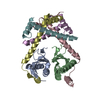
- PDB-8c26: ParDE2 toxin-antitoxin complex from Mycobacterium tuberculosis (r... -
+ Open data

- Basic information
| Entry | Database: PDB / ID: 8c26 | ||||||
|---|---|---|---|---|---|---|---|
| Title | ParDE2 toxin-antitoxin complex from Mycobacterium tuberculosis (rv2142A-rv2142c) | ||||||
 Components Components |
| ||||||
 Keywords Keywords | TOXIN / Toxin-antitoxin Gyrase inhibitor Tuberculosis | ||||||
| Function / homology |  Function and homology information Function and homology informationdetoxification / symbiont-mediated perturbation of host process / toxin sequestering activity Similarity search - Function | ||||||
| Biological species |  Mycobacterium tuberculosis H37Rv (bacteria) Mycobacterium tuberculosis H37Rv (bacteria) | ||||||
| Method |  X-RAY DIFFRACTION / SYNCHROTRON / MOLECULAR REPLACEMENT / Resolution: 2.35 Å X-RAY DIFFRACTION / SYNCHROTRON / MOLECULAR REPLACEMENT / Resolution: 2.35 Å | ||||||
 Authors Authors | Beck, I.N. / Blower, T.R. | ||||||
| Funding support |  United Kingdom, 1items United Kingdom, 1items
| ||||||
 Citation Citation | Journal: Nucleic Acids Res. / Year: 2024 Title: Toxin release by conditional remodelling of ParDE1 from Mycobacterium tuberculosis leads to gyrase inhibition. Authors: Beck, I.N. / Arrowsmith, T.J. / Grobbelaar, M.J. / Bromley, E.H.C. / Marles-Wright, J. / Blower, T.R. | ||||||
| History |
|
- Structure visualization
| Structure viewer | Molecule: MolmilJmol/JSmol |
|---|
- Downloads & links
-Download
| PDBx/mmCIF format | 8c26.cif.gz | 43 KB | Display | PDBx/mmCIF format |
|---|---|---|---|---|
| PDB format | pdb8c26.ent.gz | 26 KB | Display | PDB format |
| PDBx/mmJSON format | 8c26.json.gz | Tree view | PDBx/mmJSON format | |
| Others |  Other downloads Other downloads |
-Validation report
| Arichive directory | https://data.pdbj.org/pub/pdb/validation_reports/c2/8c26ftp://data.pdbj.org/pub/pdb/validation_reports/c2/8c26 | HTTPS FTP |
|---|
-Related structure data
-Links
 PDBj
PDBj- Assembly
| Deposited unit | 
| ||||||||||||
|---|---|---|---|---|---|---|---|---|---|---|---|---|---|
| 1 |
| ||||||||||||
| Unit cell |
| ||||||||||||
| Components on special symmetry positions |
|
-Components
| #1: Protein | Mass: 12329.859 Da / Num. of mol.: 1 Source method: isolated from a genetically manipulated source Source: (gene. exp.) Mycobacterium tuberculosis H37Rv (bacteria)Gene: parE2, Rv2142c / Production host: | ||
|---|---|---|---|
| #2: Protein | Mass: 7922.755 Da / Num. of mol.: 1 Source method: isolated from a genetically manipulated source Source: (gene. exp.) Mycobacterium tuberculosis H37Rv (bacteria)Gene: parD2, Rv2142A / Production host: | ||
| #3: Chemical |   Mass: 35.453 Da / Num. of mol.: 2 / Source method: obtained synthetically / Formula: Cl Mass: 35.453 Da / Num. of mol.: 2 / Source method: obtained synthetically / Formula: ClHas ligand of interest | N | |
-Experimental details
-Experiment
| Experiment | Method: X-RAY DIFFRACTION / Number of used crystals: 1 |
|---|
- Sample preparation
| Crystal | Density Matthews: 2.17 Å3/Da / Density % sol: 43.3 % |
|---|---|
| Crystal grow | Temperature: 294 K / Method: vapor diffusion, sitting drop / Details: 15 % PEG 3350 0.1 M MES pH 6.2 |
-Data collection
| Diffraction | Mean temperature: 100 K / Serial crystal experiment: N |
|---|---|
| Diffraction source | Source: SYNCHROTRON / Site: Diamond / Beamline: I04 / Wavelength: 0.9793 Å |
| Detector | Type: DECTRIS EIGER2 X 16M / Detector: PIXEL / Date: Oct 18, 2021 |
| Radiation | Protocol: SINGLE WAVELENGTH / Monochromatic (M) / Laue (L): M / Scattering type: x-ray |
| Radiation wavelength | Wavelength: 0.9793 Å / Relative weight: 1 |
| Reflection | Resolution: 2.345→50.59 Å / Num. obs: 7244 / % possible obs: 94.06 % / Redundancy: 19.8 % / Biso Wilson estimate: 99.74 Å2 / CC1/2: 1 / Net I/σ(I): 7.5 |
| Reflection shell | Resolution: 2.345→2.429 Å / Num. unique obs: 349 / CC1/2: 0.6 |
- Processing
| Software |
| ||||||||||||||||||||||||||||||||||||||||||
|---|---|---|---|---|---|---|---|---|---|---|---|---|---|---|---|---|---|---|---|---|---|---|---|---|---|---|---|---|---|---|---|---|---|---|---|---|---|---|---|---|---|---|---|
| Refinement | Method to determine structure: MOLECULAR REPLACEMENT / Resolution: 2.35→50.59 Å / SU ML: 0.5185 / Cross valid method: FREE R-VALUE / σ(F): 1.32 / Phase error: 41.9532 / Stereochemistry target values: CDL v1.2
| ||||||||||||||||||||||||||||||||||||||||||
| Solvent computation | Shrinkage radii: 0.9 Å / VDW probe radii: 1.11 Å / Solvent model: FLAT BULK SOLVENT MODEL | ||||||||||||||||||||||||||||||||||||||||||
| Displacement parameters | Biso mean: 102.12 Å2 | ||||||||||||||||||||||||||||||||||||||||||
| Refinement step | Cycle: LAST / Resolution: 2.35→50.59 Å
| ||||||||||||||||||||||||||||||||||||||||||
| Refine LS restraints |
| ||||||||||||||||||||||||||||||||||||||||||
| LS refinement shell |
|
