 Movie
Movie Controller

[English] 日本語
 Yorodumi
- PDB-8byh: Crystal structure of TrmD domain from Calditerrivibrio nitroreduc... -
+ Open data

- Basic information
| Entry | Database: PDB / ID: 8byh | |||||||||
|---|---|---|---|---|---|---|---|---|---|---|
| Title | Crystal structure of TrmD domain from Calditerrivibrio nitroreducens in complex with S-adenosyl-L-methionine | |||||||||
 Components Components | tRNA (guanine-N(1)-)-methyltransferase | |||||||||
 Keywords Keywords | TRANSFERASE / knotted-protein / tRNA / trmD / methyl transferase | |||||||||
| Function / homology |  Function and homology information Function and homology informationtRNA (guanine37-N1)-methyltransferase / tRNA (guanine(37)-N1)-methyltransferase activity / tRNA N1-guanine methylation / metal ion binding / cytosol Similarity search - Function | |||||||||
| Biological species |  Calditerrivibrio nitroreducens DSM 19672 (bacteria) Calditerrivibrio nitroreducens DSM 19672 (bacteria) | |||||||||
| Method |  X-RAY DIFFRACTION / SYNCHROTRON / MOLECULAR REPLACEMENT / Resolution: 2.19 Å X-RAY DIFFRACTION / SYNCHROTRON / MOLECULAR REPLACEMENT / Resolution: 2.19 Å | |||||||||
 Authors Authors | Kluza, A. / Lewandowska, I. / Sulkowska, J. | |||||||||
| Funding support |  Poland, European Union, 2items Poland, European Union, 2items
| |||||||||
 Citation Citation | Journal: To Be Published Title: Are there double knots in proteins? Prediction and in vitro verification based on TrmD-Tm1570 fusion from C. nitroreducens. To be published Authors: Sulkowska, J. | |||||||||
| History |
|
- Structure visualization
| Structure viewer | Molecule: MolmilJmol/JSmol |
|---|
- Downloads & links
-Download
| PDBx/mmCIF format | 8byh.cif.gz | 244.9 KB | Display | PDBx/mmCIF format |
|---|---|---|---|---|
| PDB format | pdb8byh.ent.gz | 172.4 KB | Display | PDB format |
| PDBx/mmJSON format | 8byh.json.gz | Tree view | PDBx/mmJSON format | |
| Others |  Other downloads Other downloads |
-Validation report
| Summary document | 8byh_validation.pdf.gz | 1 MB | Display | wwPDB validaton report |
|---|---|---|---|---|
| Full document | 8byh_full_validation.pdf.gz | 1 MB | Display | |
| Data in XML | 8byh_validation.xml.gz | 23.6 KB | Display | |
| Data in CIF | 8byh_validation.cif.gz | 34.5 KB | Display | |
| Arichive directory | https://data.pdbj.org/pub/pdb/validation_reports/by/8byhftp://data.pdbj.org/pub/pdb/validation_reports/by/8byh | HTTPS FTP |
-Related structure data
| Related structure data | 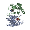 8ri0C  8b1nS S: Starting model for refinement C: citing same article ( |
|---|---|
| Similar structure data |
-Links
 PDBj
PDBj
- Assembly
| Deposited unit | 
| ||||||||||||
|---|---|---|---|---|---|---|---|---|---|---|---|---|---|
| 1 |
| ||||||||||||
| Unit cell |
| ||||||||||||
| Components on special symmetry positions |
|
-Components
-Protein , 1 types, 2 molecules AB
| #1: Protein | Mass: 27878.799 Da / Num. of mol.: 2 Source method: isolated from a genetically manipulated source Source: (gene. exp.) Calditerrivibrio nitroreducens DSM 19672 (bacteria)Gene: trmD / Production host: |
|---|
-Non-polymers , 5 types, 407 molecules 


| #2: Chemical | ChemComp-EDO /  Mass: 62.068 Da / Num. of mol.: 5 / Source method: obtained synthetically / Formula: C2H6O2 Mass: 62.068 Da / Num. of mol.: 5 / Source method: obtained synthetically / Formula: C2H6O2#3: Chemical | ChemComp-GOL / |  Mass: 92.094 Da / Num. of mol.: 1 / Source method: obtained synthetically / Formula: C3H8O3 Mass: 92.094 Da / Num. of mol.: 1 / Source method: obtained synthetically / Formula: C3H8O3#4: Chemical |  Mass: 35.453 Da / Num. of mol.: 3 / Source method: obtained synthetically / Formula: Cl Mass: 35.453 Da / Num. of mol.: 3 / Source method: obtained synthetically / Formula: Cl#5: Chemical |  Mass: 398.437 Da / Num. of mol.: 2 / Source method: obtained synthetically / Formula: C15H22N6O5S / Feature type: SUBJECT OF INVESTIGATION Mass: 398.437 Da / Num. of mol.: 2 / Source method: obtained synthetically / Formula: C15H22N6O5S / Feature type: SUBJECT OF INVESTIGATION#6: Water | ChemComp-HOH / | Mass: 18.015 Da / Num. of mol.: 396 / Source method: isolated from a natural source / Formula: H2O |
|---|
-Details
| Has ligand of interest | Y |
|---|
-Experimental details
-Experiment
| Experiment | Method: X-RAY DIFFRACTION / Number of used crystals: 1 |
|---|
- Sample preparation
| Crystal | Density Matthews: 3.51 Å3/Da / Density % sol: 65 % |
|---|---|
| Crystal grow | Temperature: 293 K / Method: vapor diffusion, sitting drop Details: Protein sample: 4 mg/mL TrmD in 50 mM HEPES buffer pH 7.4, 300 mM NaCl, 5% glycerol, 10 mM SAM, 20 mM MgCl2. Reservoir solution: 0.2 M sodium thiocyanate, 20% w/v PEG 3350 (JCSG+ screen 1-14). |
-Data collection
| Diffraction | Mean temperature: 100 K / Serial crystal experiment: N |
|---|---|
| Diffraction source | Source: SYNCHROTRON / Site: PETRA III, EMBL c/o DESY  / Beamline: P14 (MX2) / Wavelength: 0.46522 Å / Beamline: P14 (MX2) / Wavelength: 0.46522 Å |
| Detector | Type: DECTRIS EIGER2 X CdTe 16M / Detector: PIXEL / Date: Oct 27, 2022 |
| Radiation | Protocol: SINGLE WAVELENGTH / Monochromatic (M) / Laue (L): M / Scattering type: x-ray |
| Radiation wavelength | Wavelength: 0.46522 Å / Relative weight: 1 |
| Reflection | Resolution: 2.19→45.79 Å / Num. obs: 40840 / % possible obs: 99.1 % / Redundancy: 8.75 % / Biso Wilson estimate: 32.88 Å2 / CC1/2: 0.998 / Rrim(I) all: 0.133 / Net I/σ(I): 11.93 |
| Reflection shell | Resolution: 2.19→2.32 Å / Redundancy: 5.67 % / Mean I/σ(I) obs: 1.8 / Num. unique obs: 6191 / CC1/2: 0.72 / Rrim(I) all: 1.018 / % possible all: 95.1 |
- Processing
| Software |
| |||||||||||||||||||||||||||||||||||||||||||||||||||||||||||||||||||||||||||||||||||||||||||||||||||||||||||||||||||||||||||||||||||||||||||||||||||||||||||||||||||||||||||||||
|---|---|---|---|---|---|---|---|---|---|---|---|---|---|---|---|---|---|---|---|---|---|---|---|---|---|---|---|---|---|---|---|---|---|---|---|---|---|---|---|---|---|---|---|---|---|---|---|---|---|---|---|---|---|---|---|---|---|---|---|---|---|---|---|---|---|---|---|---|---|---|---|---|---|---|---|---|---|---|---|---|---|---|---|---|---|---|---|---|---|---|---|---|---|---|---|---|---|---|---|---|---|---|---|---|---|---|---|---|---|---|---|---|---|---|---|---|---|---|---|---|---|---|---|---|---|---|---|---|---|---|---|---|---|---|---|---|---|---|---|---|---|---|---|---|---|---|---|---|---|---|---|---|---|---|---|---|---|---|---|---|---|---|---|---|---|---|---|---|---|---|---|---|---|---|---|---|
| Refinement | Method to determine structure: MOLECULAR REPLACEMENT Starting model: 8B1N Resolution: 2.19→44.83 Å / SU ML: 0.2483 / Cross valid method: FREE R-VALUE / σ(F): 1.34 / Phase error: 21.8238 Stereochemistry target values: GeoStd + Monomer Library + CDL v1.2
| |||||||||||||||||||||||||||||||||||||||||||||||||||||||||||||||||||||||||||||||||||||||||||||||||||||||||||||||||||||||||||||||||||||||||||||||||||||||||||||||||||||||||||||||
| Solvent computation | Shrinkage radii: 0.9 Å / VDW probe radii: 1.11 Å / Solvent model: FLAT BULK SOLVENT MODEL | |||||||||||||||||||||||||||||||||||||||||||||||||||||||||||||||||||||||||||||||||||||||||||||||||||||||||||||||||||||||||||||||||||||||||||||||||||||||||||||||||||||||||||||||
| Displacement parameters | Biso mean: 40.66 Å2 | |||||||||||||||||||||||||||||||||||||||||||||||||||||||||||||||||||||||||||||||||||||||||||||||||||||||||||||||||||||||||||||||||||||||||||||||||||||||||||||||||||||||||||||||
| Refinement step | Cycle: LAST / Resolution: 2.19→44.83 Å
| |||||||||||||||||||||||||||||||||||||||||||||||||||||||||||||||||||||||||||||||||||||||||||||||||||||||||||||||||||||||||||||||||||||||||||||||||||||||||||||||||||||||||||||||
| Refine LS restraints |
| |||||||||||||||||||||||||||||||||||||||||||||||||||||||||||||||||||||||||||||||||||||||||||||||||||||||||||||||||||||||||||||||||||||||||||||||||||||||||||||||||||||||||||||||
| LS refinement shell |
| |||||||||||||||||||||||||||||||||||||||||||||||||||||||||||||||||||||||||||||||||||||||||||||||||||||||||||||||||||||||||||||||||||||||||||||||||||||||||||||||||||||||||||||||
| Refinement TLS params. | Method: refined / Refine-ID: X-RAY DIFFRACTION
| |||||||||||||||||||||||||||||||||||||||||||||||||||||||||||||||||||||||||||||||||||||||||||||||||||||||||||||||||||||||||||||||||||||||||||||||||||||||||||||||||||||||||||||||
| Refinement TLS group |
|
