 Movie
Movie Controller

[English] 日本語
 Yorodumi
- PDB-8bqg: W-formate dehydrogenase from Desulfovibrio vulgaris - Soaking wit... -
+ Open data

- Basic information
| Entry | Database: PDB / ID: 8bqg | ||||||||||||||||||
|---|---|---|---|---|---|---|---|---|---|---|---|---|---|---|---|---|---|---|---|
| Title | W-formate dehydrogenase from Desulfovibrio vulgaris - Soaking with Formate 1 min | ||||||||||||||||||
 Components Components | (Formate dehydrogenase, ...) x 2 | ||||||||||||||||||
 Keywords Keywords | OXIDOREDUCTASE / Formate / CO2 / Molybdenum and Tungsten enzymes / DMSO reductase family | ||||||||||||||||||
| Function / homology |  Function and homology information Function and homology informationformate dehydrogenase (cytochrome-c-553) activity / formate dehydrogenase / formate dehydrogenase (NAD+) activity / molybdenum ion binding / cell envelope / molybdopterin cofactor binding / anaerobic respiration / 4 iron, 4 sulfur cluster binding / electron transfer activity / periplasmic space / metal ion binding Similarity search - Function | ||||||||||||||||||
| Biological species |  Desulfovibrio vulgaris str. Hildenborough (bacteria) Desulfovibrio vulgaris str. Hildenborough (bacteria) | ||||||||||||||||||
| Method |  X-RAY DIFFRACTION / SYNCHROTRON / MOLECULAR REPLACEMENT / Resolution: 1.946 Å X-RAY DIFFRACTION / SYNCHROTRON / MOLECULAR REPLACEMENT / Resolution: 1.946 Å | ||||||||||||||||||
 Authors Authors | Vilela-Alves, G. / Mota, C. / Oliveira, A.R. / Manuel, R.R. / Pereira, I.C. / Romao, M.J. | ||||||||||||||||||
| Funding support |  Portugal, 5items Portugal, 5items
| ||||||||||||||||||
 Citation Citation | Journal: Int J Mol Sci / Year: 2022 Title: Tracking W-Formate Dehydrogenase Structural Changes During Catalysis and Enzyme Reoxidation. Authors: Vilela-Alves, G. / Manuel, R.R. / Oliveira, A.R. / Pereira, I.C. / Romao, M.J. / Mota, C. | ||||||||||||||||||
| History |
|
- Structure visualization
| Structure viewer | Molecule: MolmilJmol/JSmol |
|---|
- Downloads & links
-Download
| PDBx/mmCIF format | 8bqg.cif.gz | 264.8 KB | Display | PDBx/mmCIF format |
|---|---|---|---|---|
| PDB format | pdb8bqg.ent.gz | 197.2 KB | Display | PDB format |
| PDBx/mmJSON format | 8bqg.json.gz | Tree view | PDBx/mmJSON format | |
| Others |  Other downloads Other downloads |
-Validation report
| Arichive directory | https://data.pdbj.org/pub/pdb/validation_reports/bq/8bqgftp://data.pdbj.org/pub/pdb/validation_reports/bq/8bqg | HTTPS FTP |
|---|
-Related structure data
| Related structure data |  8bqhC  8bqiC  8bqjC  8bqkC  8bqlC 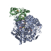 6sdrS S: Starting model for refinement C: citing same article ( |
|---|---|
| Similar structure data | |
| Experimental dataset #1 | Data reference: 10.15151/ESRF-ES-643877202 / Data set type: diffraction image data |
-Links
 PDBj
PDBj


- Assembly
| Deposited unit | 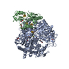
| ||||||||
|---|---|---|---|---|---|---|---|---|---|
| 1 |
| ||||||||
| Unit cell |
|
-Components
-Formate dehydrogenase, ... , 2 types, 2 molecules AB
| #1: Protein | Mass: 112437.023 Da / Num. of mol.: 1 Source method: isolated from a genetically manipulated source Source: (gene. exp.) Desulfovibrio vulgaris str. Hildenborough (bacteria)Strain: Hildenborough / Gene: fdnG-1 Production host: Desulfovibrio vulgaris str. Hildenborough (bacteria)Strain (production host): Hildenborough / References: UniProt: Q72EJ1 |
|---|---|
| #2: Protein | Mass: 26481.494 Da / Num. of mol.: 1 Source method: isolated from a genetically manipulated source Source: (gene. exp.) Desulfovibrio vulgaris str. Hildenborough (bacteria)Strain: Hildenborough / Gene: DVU_0588 Production host: Desulfovibrio vulgaris str. Hildenborough (bacteria)Strain (production host): Hildenborough / References: UniProt: Q72EJ0 |
-Non-polymers , 9 types, 163 molecules 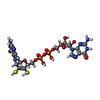

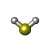


| #3: Chemical |  Mass: 740.557 Da / Num. of mol.: 2 / Source method: obtained synthetically / Formula: C20H26N10O13P2S2 / Feature type: SUBJECT OF INVESTIGATION Mass: 740.557 Da / Num. of mol.: 2 / Source method: obtained synthetically / Formula: C20H26N10O13P2S2 / Feature type: SUBJECT OF INVESTIGATION#4: Chemical | ChemComp-SF4 /  Mass: 351.640 Da / Num. of mol.: 4 / Source method: obtained synthetically / Formula: Fe4S4 / Feature type: SUBJECT OF INVESTIGATION Mass: 351.640 Da / Num. of mol.: 4 / Source method: obtained synthetically / Formula: Fe4S4 / Feature type: SUBJECT OF INVESTIGATION#5: Chemical | ChemComp-H2S / |  Mass: 34.081 Da / Num. of mol.: 1 / Source method: obtained synthetically / Formula: H2S / Feature type: SUBJECT OF INVESTIGATION Mass: 34.081 Da / Num. of mol.: 1 / Source method: obtained synthetically / Formula: H2S / Feature type: SUBJECT OF INVESTIGATION#6: Chemical | ChemComp-W / |  Mass: 183.840 Da / Num. of mol.: 1 / Source method: obtained synthetically / Formula: W / Feature type: SUBJECT OF INVESTIGATION Mass: 183.840 Da / Num. of mol.: 1 / Source method: obtained synthetically / Formula: W / Feature type: SUBJECT OF INVESTIGATION#7: Chemical |  Mass: 92.094 Da / Num. of mol.: 3 / Source method: obtained synthetically / Formula: C3H8O3 Mass: 92.094 Da / Num. of mol.: 3 / Source method: obtained synthetically / Formula: C3H8O3#8: Chemical | ChemComp-PEG / |  Mass: 106.120 Da / Num. of mol.: 1 / Source method: obtained synthetically / Formula: C4H10O3 Mass: 106.120 Da / Num. of mol.: 1 / Source method: obtained synthetically / Formula: C4H10O3#9: Chemical |  Mass: 62.068 Da / Num. of mol.: 2 / Source method: obtained synthetically / Formula: C2H6O2 Mass: 62.068 Da / Num. of mol.: 2 / Source method: obtained synthetically / Formula: C2H6O2#10: Chemical | ChemComp-FMT / |  Mass: 46.025 Da / Num. of mol.: 1 / Source method: obtained synthetically / Formula: CH2O2 / Feature type: SUBJECT OF INVESTIGATION Mass: 46.025 Da / Num. of mol.: 1 / Source method: obtained synthetically / Formula: CH2O2 / Feature type: SUBJECT OF INVESTIGATION#11: Water | ChemComp-HOH / | Mass: 18.015 Da / Num. of mol.: 148 / Source method: isolated from a natural source / Formula: H2O |
|---|
-Details
| Has ligand of interest | Y |
|---|---|
| Has protein modification | Y |
-Experimental details
-Experiment
| Experiment | Method: X-RAY DIFFRACTION / Number of used crystals: 1 |
|---|
- Sample preparation
| Crystal | Density Matthews: 2.24 Å3/Da / Density % sol: 44.98 % |
|---|---|
| Crystal grow | Temperature: 293 K / Method: vapor diffusion, hanging drop / pH: 8 / Details: 24% PEG 3350, 0.1M Tris-HCl pH 8.0, 1M LiCl |
-Data collection
| Diffraction | Mean temperature: 100 K / Serial crystal experiment: N |
|---|---|
| Diffraction source | Source: SYNCHROTRON / Site: ESRF  / Beamline: ID23-1 / Wavelength: 0.8856 Å / Beamline: ID23-1 / Wavelength: 0.8856 Å |
| Detector | Type: DECTRIS EIGER2 S 16M / Detector: PIXEL / Date: Mar 1, 2022 |
| Radiation | Protocol: SINGLE WAVELENGTH / Monochromatic (M) / Laue (L): M / Scattering type: x-ray |
| Radiation wavelength | Wavelength: 0.8856 Å / Relative weight: 1 |
| Reflection | Resolution: 1.946→94.93 Å / Num. obs: 73334 / % possible obs: 91.96 % / Redundancy: 4.33 % / CC1/2: 0.9971 / Net I/σ(I): 9.2 |
| Reflection shell | Resolution: 1.95→2.07 Å / Num. unique obs: 3667 / CC1/2: 0.435 |
- Processing
| Software |
| |||||||||||||||||||||||||||||||||||||||||||||||||||||||||||||||||||||||||||||||||||||||||||||||||||||||||||||||||||||||||||||||||||||||||||||||||||||||||||
|---|---|---|---|---|---|---|---|---|---|---|---|---|---|---|---|---|---|---|---|---|---|---|---|---|---|---|---|---|---|---|---|---|---|---|---|---|---|---|---|---|---|---|---|---|---|---|---|---|---|---|---|---|---|---|---|---|---|---|---|---|---|---|---|---|---|---|---|---|---|---|---|---|---|---|---|---|---|---|---|---|---|---|---|---|---|---|---|---|---|---|---|---|---|---|---|---|---|---|---|---|---|---|---|---|---|---|---|---|---|---|---|---|---|---|---|---|---|---|---|---|---|---|---|---|---|---|---|---|---|---|---|---|---|---|---|---|---|---|---|---|---|---|---|---|---|---|---|---|---|---|---|---|---|---|---|---|
| Refinement | Method to determine structure: MOLECULAR REPLACEMENT Starting model: 6SDR Resolution: 1.946→94.93 Å / Cor.coef. Fo:Fc: 0.962 / Cor.coef. Fo:Fc free: 0.938 / WRfactor Rfree: 0.232 / WRfactor Rwork: 0.178 / SU B: 6.04 / SU ML: 0.159 / Average fsc free: 0.8701 / Average fsc work: 0.8885 / Cross valid method: FREE R-VALUE / ESU R: 0.222 / ESU R Free: 0.188 Details: Hydrogens have been added in their riding positions
| |||||||||||||||||||||||||||||||||||||||||||||||||||||||||||||||||||||||||||||||||||||||||||||||||||||||||||||||||||||||||||||||||||||||||||||||||||||||||||
| Solvent computation | Ion probe radii: 0.8 Å / Shrinkage radii: 0.8 Å / VDW probe radii: 1.2 Å / Solvent model: MASK BULK SOLVENT | |||||||||||||||||||||||||||||||||||||||||||||||||||||||||||||||||||||||||||||||||||||||||||||||||||||||||||||||||||||||||||||||||||||||||||||||||||||||||||
| Displacement parameters | Biso mean: 38.302 Å2
| |||||||||||||||||||||||||||||||||||||||||||||||||||||||||||||||||||||||||||||||||||||||||||||||||||||||||||||||||||||||||||||||||||||||||||||||||||||||||||
| Refinement step | Cycle: LAST / Resolution: 1.946→94.93 Å
| |||||||||||||||||||||||||||||||||||||||||||||||||||||||||||||||||||||||||||||||||||||||||||||||||||||||||||||||||||||||||||||||||||||||||||||||||||||||||||
| Refine LS restraints |
| |||||||||||||||||||||||||||||||||||||||||||||||||||||||||||||||||||||||||||||||||||||||||||||||||||||||||||||||||||||||||||||||||||||||||||||||||||||||||||
| LS refinement shell |
|
