 Movie
Movie Controller


+ Open data

- Basic information
| Entry | Database: PDB / ID: 8bn0 | ||||||
|---|---|---|---|---|---|---|---|
| Title | Bacteroides thetaiotaomicron surface protein BT1954 bound to | ||||||
 Components Components | Putative surface layer protein | ||||||
 Keywords Keywords | MEMBRANE PROTEIN / Surface exposed protein Beta propeller Vitamin B12 binding protein | ||||||
| Function / homology |  Function and homology information Function and homology informationProtein of unknown function DUF5074 / Domain of unknown function (DUF5074) / : / Cytochrome cd1-nitrite reductase-like, haem d1 domain superfamily / YVTN repeat-like/Quinoprotein amine dehydrogenase / 7 Propeller / Methylamine Dehydrogenase; Chain H / Prokaryotic membrane lipoprotein lipid attachment site profile. / WD40/YVTN repeat-like-containing domain superfamily / Mainly Beta Similarity search - Domain/homology | ||||||
| Biological species |  Bacteroides thetaiotaomicron VPI-5482 (bacteria) Bacteroides thetaiotaomicron VPI-5482 (bacteria) | ||||||
| Method |  X-RAY DIFFRACTION / SYNCHROTRON / MOLECULAR REPLACEMENT / Resolution: 1.33 Å X-RAY DIFFRACTION / SYNCHROTRON / MOLECULAR REPLACEMENT / Resolution: 1.33 Å | ||||||
 Authors Authors | Abellon-Ruiz, J. / Jana, K. / Silale, A. / Basle, A. / Kleinekathofer, U. / van den Berg, B. | ||||||
| Funding support |  United Kingdom, 1items United Kingdom, 1items
| ||||||
 Citation Citation | Journal: Nat Commun / Year: 2023 Title: BtuB TonB-dependent transporters and BtuG surface lipoproteins form stable complexes for vitamin B uptake in gut Bacteroides. Authors: Javier Abellon-Ruiz / Kalyanashis Jana / Augustinas Silale / Andrew M Frey / Arnaud Baslé / Matthias Trost / Ulrich Kleinekathöfer / Bert van den Berg /  Abstract: Vitamin B (cobalamin) is required for most human gut microbes, many of which are dependent on scavenging to obtain this vitamin. Since bacterial densities in the gut are extremely high, competition ...Vitamin B (cobalamin) is required for most human gut microbes, many of which are dependent on scavenging to obtain this vitamin. Since bacterial densities in the gut are extremely high, competition for this keystone micronutrient is severe. Contrasting with Enterobacteria, members of the dominant genus Bacteroides often encode several BtuB vitamin B outer membrane transporters together with a conserved array of surface-exposed B-binding lipoproteins. Here we show that the BtuB transporters from Bacteroides thetaiotaomicron form stable, pedal bin-like complexes with surface-exposed BtuG lipoprotein lids, which bind B with high affinities. Closing of the BtuG lid following B capture causes destabilisation of the bound B by a conserved BtuB extracellular loop, causing translocation of the vitamin to BtuB and subsequent transport. We propose that TonB-dependent, lipoprotein-assisted small molecule uptake is a general feature of Bacteroides spp. that is important for the success of this genus in colonising the human gut. | ||||||
| History |
|
- Structure visualization
| Structure viewer | Molecule: MolmilJmol/JSmol |
|---|
- Downloads & links
-Download
| PDBx/mmCIF format | 8bn0.cif.gz | 209.9 KB | Display | PDBx/mmCIF format |
|---|---|---|---|---|
| PDB format | pdb8bn0.ent.gz | 137.3 KB | Display | PDB format |
| PDBx/mmJSON format | 8bn0.json.gz | Tree view | PDBx/mmJSON format | |
| Others |  Other downloads Other downloads |
-Validation report
| Arichive directory | https://data.pdbj.org/pub/pdb/validation_reports/bn/8bn0ftp://data.pdbj.org/pub/pdb/validation_reports/bn/8bn0 | HTTPS FTP |
|---|
-Related structure data
| Related structure data |  8blwC  8bmxC 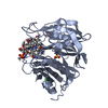 8bmyC 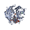 8bmzC 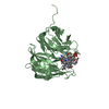 8okvC 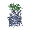 8p97C 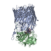 8p98C  3dsmS S: Starting model for refinement C: citing same article ( |
|---|---|
| Similar structure data |


-Links
 PDBj
PDBj


- Assembly
| Deposited unit | 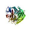
| ||||||||||||
|---|---|---|---|---|---|---|---|---|---|---|---|---|---|
| 1 |
| ||||||||||||
| Unit cell |
|
-Components
| #1: Protein | Mass: 43632.234 Da / Num. of mol.: 1 Source method: isolated from a genetically manipulated source Source: (gene. exp.) Bacteroides thetaiotaomicron VPI-5482 (bacteria)Strain: ATCC 29148 / DSM 2079 / JCM 5827 / CCUG 10774 / NCTC 10582 / VPI-5482 / E50 Gene: BT_1954 / Production host: |
|---|---|
| #2: Chemical | ChemComp-CYN /   Mass: 26.017 Da / Num. of mol.: 1 / Source method: obtained synthetically / Formula: CN Mass: 26.017 Da / Num. of mol.: 1 / Source method: obtained synthetically / Formula: CN |
| #3: Chemical | ChemComp-CL /   Mass: 35.453 Da / Num. of mol.: 1 / Source method: obtained synthetically / Formula: Cl Mass: 35.453 Da / Num. of mol.: 1 / Source method: obtained synthetically / Formula: Cl |
| #4: Chemical | ChemComp-CBY / 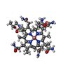  Mass: 990.087 Da / Num. of mol.: 1 / Source method: obtained synthetically / Formula: C48H72CoN11O8 / Feature type: SUBJECT OF INVESTIGATION Mass: 990.087 Da / Num. of mol.: 1 / Source method: obtained synthetically / Formula: C48H72CoN11O8 / Feature type: SUBJECT OF INVESTIGATION |
| #5: Water | ChemComp-HOH /  Mass: 18.015 Da / Num. of mol.: 414 / Source method: isolated from a natural source / Formula: H2O Mass: 18.015 Da / Num. of mol.: 414 / Source method: isolated from a natural source / Formula: H2O |
| Has ligand of interest | Y |
| Has protein modification | N |
-Experimental details
-Experiment
| Experiment | Method: X-RAY DIFFRACTION / Number of used crystals: 1 |
|---|
- Sample preparation
| Crystal | Density Matthews: 2.78 Å3/Da / Density % sol: 55.74 % |
|---|---|
| Crystal grow | Temperature: 293 K / Method: vapor diffusion, sitting drop Details: 1.1M Na malonate pH7.0 0.1M HEPES pH 7.0 0.5% Jefamine ED2001 pH7 |
-Data collection
| Diffraction | Mean temperature: 100 K / Serial crystal experiment: N |
|---|---|
| Diffraction source | Source: SYNCHROTRON / Site: Diamond / Beamline: I24 / Wavelength: 0.9998 Å |
| Detector | Type: DECTRIS PILATUS3 6M / Detector: PIXEL / Date: Feb 26, 2022 |
| Radiation | Protocol: SINGLE WAVELENGTH / Monochromatic (M) / Laue (L): M / Scattering type: x-ray |
| Radiation wavelength | Wavelength: 0.9998 Å / Relative weight: 1 |
| Reflection | Resolution: 1.33→43.3 Å / Num. obs: 113086 / % possible obs: 99.85 % / Redundancy: 24.2 % / Biso Wilson estimate: 18.53 Å2 / CC1/2: 1 / Net I/σ(I): 16.7 |
| Reflection shell | Resolution: 1.33→1.36 Å / Mean I/σ(I) obs: 0.9 / Num. unique obs: 11000 / CC1/2: 0.3 |
- Processing
| Software |
| |||||||||||||||||||||||||||||||||||||||||||||||||||||||||||||||||||||||||||||||||||||||||||||||||||||||||||||||||||||||||||||||||||||||||||||||||||||||||||||||||||||||||||||||||||||||||||||||||||||||||||||||||||||||||
|---|---|---|---|---|---|---|---|---|---|---|---|---|---|---|---|---|---|---|---|---|---|---|---|---|---|---|---|---|---|---|---|---|---|---|---|---|---|---|---|---|---|---|---|---|---|---|---|---|---|---|---|---|---|---|---|---|---|---|---|---|---|---|---|---|---|---|---|---|---|---|---|---|---|---|---|---|---|---|---|---|---|---|---|---|---|---|---|---|---|---|---|---|---|---|---|---|---|---|---|---|---|---|---|---|---|---|---|---|---|---|---|---|---|---|---|---|---|---|---|---|---|---|---|---|---|---|---|---|---|---|---|---|---|---|---|---|---|---|---|---|---|---|---|---|---|---|---|---|---|---|---|---|---|---|---|---|---|---|---|---|---|---|---|---|---|---|---|---|---|---|---|---|---|---|---|---|---|---|---|---|---|---|---|---|---|---|---|---|---|---|---|---|---|---|---|---|---|---|---|---|---|---|---|---|---|---|---|---|---|---|---|---|---|---|---|---|---|---|
| Refinement | Method to determine structure: MOLECULAR REPLACEMENT Starting model: 3DSM Resolution: 1.33→43.3 Å / SU ML: 0.1523 / Cross valid method: FREE R-VALUE / σ(F): 1.34 / Phase error: 17.1931 Stereochemistry target values: GeoStd + Monomer Library + CDL v1.2
| |||||||||||||||||||||||||||||||||||||||||||||||||||||||||||||||||||||||||||||||||||||||||||||||||||||||||||||||||||||||||||||||||||||||||||||||||||||||||||||||||||||||||||||||||||||||||||||||||||||||||||||||||||||||||
| Solvent computation | Shrinkage radii: 0.9 Å / VDW probe radii: 1.11 Å / Solvent model: FLAT BULK SOLVENT MODEL | |||||||||||||||||||||||||||||||||||||||||||||||||||||||||||||||||||||||||||||||||||||||||||||||||||||||||||||||||||||||||||||||||||||||||||||||||||||||||||||||||||||||||||||||||||||||||||||||||||||||||||||||||||||||||
| Displacement parameters | Biso mean: 25.2 Å2 | |||||||||||||||||||||||||||||||||||||||||||||||||||||||||||||||||||||||||||||||||||||||||||||||||||||||||||||||||||||||||||||||||||||||||||||||||||||||||||||||||||||||||||||||||||||||||||||||||||||||||||||||||||||||||
| Refinement step | Cycle: LAST / Resolution: 1.33→43.3 Å
| |||||||||||||||||||||||||||||||||||||||||||||||||||||||||||||||||||||||||||||||||||||||||||||||||||||||||||||||||||||||||||||||||||||||||||||||||||||||||||||||||||||||||||||||||||||||||||||||||||||||||||||||||||||||||
| Refine LS restraints |
| |||||||||||||||||||||||||||||||||||||||||||||||||||||||||||||||||||||||||||||||||||||||||||||||||||||||||||||||||||||||||||||||||||||||||||||||||||||||||||||||||||||||||||||||||||||||||||||||||||||||||||||||||||||||||
| LS refinement shell |
|
