| Entry | Database: PDB / ID: 8bjk
|
|---|
| Title | X-ray structure of Danio rerio histone deacetylase 6 (HDAC6) CD2 in complex with an inhibitor CPD11352 |
|---|
 Components Components | Histone deacetylase 6 |
|---|
 Keywords Keywords | HYDROLASE / HISTONE DEACETYLASE / DEACETYLATION / INHIBITOR / DANIO RERIO |
|---|
| Function / homology |  Function and homology information Function and homology information
Aggrephagy / negative regulation of cellular component organization / positive regulation of cellular component organization / deacetylase activity / tubulin deacetylase activity / mitochondrion localization / definitive hemopoiesis / regulation of microtubule-based process / protein lysine deacetylase activity / potassium ion binding ...Aggrephagy / negative regulation of cellular component organization / positive regulation of cellular component organization / deacetylase activity / tubulin deacetylase activity / mitochondrion localization / definitive hemopoiesis / regulation of microtubule-based process / protein lysine deacetylase activity / potassium ion binding / response to stress / hematopoietic progenitor cell differentiation / swimming behavior / transferase activity / chromatin organization / actin binding / angiogenesis / perikaryon / axon / centrosome / dendrite / zinc ion binding / nucleus / cytoplasm / cytosolSimilarity search - Function Ubiquitin Carboxyl-terminal Hydrolase-like zinc finger / Zinc finger, UBP-type / Zn-finger in ubiquitin-hydrolases and other protein / Zinc finger UBP-type profile. / Histone deacetylase family / Histone deacetylase domain / Histone deacetylase domain superfamily / Histone deacetylase domain / Ureohydrolase domain superfamily / Zinc finger, RING/FYVE/PHD-typeSimilarity search - Domain/homology |
|---|
| Biological species |   Danio rerio (zebrafish) Danio rerio (zebrafish) |
|---|
| Method |  X-RAY DIFFRACTION / FOURIER SYNTHESIS / Resolution: 1.35 Å X-RAY DIFFRACTION / FOURIER SYNTHESIS / Resolution: 1.35 Å |
|---|
 Authors Authors | Barinka, C. / Motlova, L. / Pavlicek, J. |
|---|
| Funding support | 1items | Organization | Grant number | Country |
|---|
| Not funded | | |
|
|---|
 Citation Citation | Journal: Acs Chem.Biol. / Year: 2023
Title: Comprehensive Mechanistic View of the Hydrolysis of Oxadiazole-Based Inhibitors by Histone Deacetylase 6 (HDAC6).
Authors: Motlova, L. / Snajdr, I. / Kutil, Z. / Andris, E. / Ptacek, J. / Novotna, A. / Novakova, Z. / Havlinova, B. / Tueckmantel, W. / Draberova, H. / Majer, P. / Schutkowski, M. / Kozikowski, A. / ...Authors: Motlova, L. / Snajdr, I. / Kutil, Z. / Andris, E. / Ptacek, J. / Novotna, A. / Novakova, Z. / Havlinova, B. / Tueckmantel, W. / Draberova, H. / Majer, P. / Schutkowski, M. / Kozikowski, A. / Rulisek, L. / Barinka, C. |
|---|
| History | | Deposition | Nov 4, 2022 | Deposition site: PDBE / Processing site: PDBE |
|---|
| Revision 1.0 | Sep 6, 2023 | Provider: repository / Type: Initial release |
|---|
| Revision 1.1 | Mar 4, 2026 | Group: Refinement description / Structure summary
Category: pdbx_entry_details / pdbx_initial_refinement_model
Item: _pdbx_entry_details.has_protein_modification |
|---|
|
|---|
 Movie
Movie Controller

 Yorodumi
 Open data
 Basic information
 Structure visualization
 Downloads & links
 Other downloads
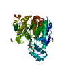
 PDBj
PDBj Assembly
 Homo sapiens (human) / References: UniProt: F8W4B7
 Mass: 65.409 Da / Num. of mol.: 1 / Source method: obtained synthetically / Formula: Zn
 Mass: 39.098 Da / Num. of mol.: 2 / Source method: obtained synthetically / Formula: K
 Mass: 354.812 Da / Num. of mol.: 1 / Source method: obtained synthetically / Formula: C14H15ClN4O3S / Feature type: SUBJECT OF INVESTIGATION
 Mass: 18.015 Da / Num. of mol.: 369 / Source method: isolated from a natural source / Formula: H2O
 Sample preparation
 Processing