 Movie
Movie Controller


+ Open data

- Basic information
| Entry | Database: PDB / ID: 8bjc | ||||||
|---|---|---|---|---|---|---|---|
| Title | Full length structure of the apo-state LpMIP. | ||||||
 Components Components | Peptidyl-prolyl cis-trans isomerase | ||||||
 Keywords Keywords | STRUCTURAL PROTEIN / Macrophage / potentiator / soluble / protein. | ||||||
| Function / homology |  Function and homology information Function and homology informationpeptidylprolyl isomerase / peptidyl-prolyl cis-trans isomerase activity / cell outer membrane / protein folding Similarity search - Function | ||||||
| Biological species |   Legionella pneumophila (bacteria) Legionella pneumophila (bacteria) | ||||||
| Method |  X-RAY DIFFRACTION / SYNCHROTRON / MOLECULAR REPLACEMENT / Resolution: 1.71 Å X-RAY DIFFRACTION / SYNCHROTRON / MOLECULAR REPLACEMENT / Resolution: 1.71 Å | ||||||
 Authors Authors | Whittaker, J.J. / Guskov, A. / Goretzki, B. / Hellmich, U.A. | ||||||
| Funding support |  Germany, 1items Germany, 1items
| ||||||
 Citation Citation | Journal: Int.J.Biol.Macromol. / Year: 2023 Title: Legionella pneumophila macrophage infectivity potentiator protein appendage domains modulate protein dynamics and inhibitor binding. Authors: Wiedemann, C. / Whittaker, J.J. / Perez Carrillo, V.H. / Goretzki, B. / Dajka, M. / Tebbe, F. / Harder, J.M. / Krajczy, P.R. / Joseph, B. / Hausch, F. / Guskov, A. / Hellmich, U.A. | ||||||
| History |
|
- Structure visualization
| Structure viewer | Molecule: MolmilJmol/JSmol |
|---|
- Downloads & links
-Download
| PDBx/mmCIF format | 8bjc.cif.gz | 93 KB | Display | PDBx/mmCIF format |
|---|---|---|---|---|
| PDB format | pdb8bjc.ent.gz | 69.7 KB | Display | PDB format |
| PDBx/mmJSON format | 8bjc.json.gz | Tree view | PDBx/mmJSON format | |
| Others |  Other downloads Other downloads |
-Validation report
| Arichive directory | https://data.pdbj.org/pub/pdb/validation_reports/bj/8bjcftp://data.pdbj.org/pub/pdb/validation_reports/bj/8bjc | HTTPS FTP |
|---|
-Related structure data
| Related structure data | 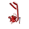 8bjdC 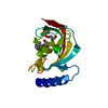 8bjeC  8bk4C 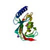 8bk5C 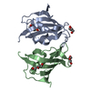 8bk6C 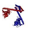 1fd9S S: Starting model for refinement C: citing same article ( |
|---|---|
| Similar structure data |
-Links
 PDBj
PDBj

- Assembly
| Deposited unit | 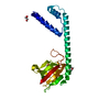
| ||||||||
|---|---|---|---|---|---|---|---|---|---|
| 1 |
| ||||||||
| Unit cell |
|
-Components
-Protein , 1 types, 1 molecules B
| #1: Protein | Mass: 22148.326 Da / Num. of mol.: 1 Source method: isolated from a genetically manipulated source Source: (gene. exp.) Legionella pneumophila (bacteria) / Gene: C3927_03235 / Production host: |
|---|
-Non-polymers , 5 types, 116 molecules 


| #2: Chemical | ChemComp-GOL /  Mass: 92.094 Da / Num. of mol.: 1 / Source method: obtained synthetically / Formula: C3H8O3 Mass: 92.094 Da / Num. of mol.: 1 / Source method: obtained synthetically / Formula: C3H8O3 | ||||||
|---|---|---|---|---|---|---|---|
| #3: Chemical |  Mass: 238.305 Da / Num. of mol.: 2 / Source method: obtained synthetically / Formula: C8H18N2O4S / Comment: pH buffer*YM Mass: 238.305 Da / Num. of mol.: 2 / Source method: obtained synthetically / Formula: C8H18N2O4S / Comment: pH buffer*YM#4: Chemical | ChemComp-MES / |  Mass: 195.237 Da / Num. of mol.: 1 / Source method: obtained synthetically / Formula: C6H13NO4S / Comment: pH buffer*YM Mass: 195.237 Da / Num. of mol.: 1 / Source method: obtained synthetically / Formula: C6H13NO4S / Comment: pH buffer*YM#5: Chemical | ChemComp-ZN / |  Mass: 65.409 Da / Num. of mol.: 1 / Source method: obtained synthetically / Formula: Zn Mass: 65.409 Da / Num. of mol.: 1 / Source method: obtained synthetically / Formula: Zn#6: Water | ChemComp-HOH / | Mass: 18.015 Da / Num. of mol.: 111 / Source method: isolated from a natural source / Formula: H2O |
-Details
| Has ligand of interest | N |
|---|---|
| Has protein modification | N |
-Experimental details
-Experiment
| Experiment | Method: X-RAY DIFFRACTION / Number of used crystals: 1 |
|---|
- Sample preparation
| Crystal | Density Matthews: 3.43 Å3/Da / Density % sol: 64.15 % |
|---|---|
| Crystal grow | Temperature: 298 K / Method: vapor diffusion, sitting drop / pH: 6 Details: 20 %(w/v) PEG 6000, 500 mM zinc acetate dihydrate, 100 mM MES, pH 6.0. |
-Data collection
| Diffraction | Mean temperature: 100 K / Serial crystal experiment: N |
|---|---|
| Diffraction source | Source: SYNCHROTRON / Site: ESRF  / Beamline: ID23-1 / Wavelength: 0.9673 Å / Beamline: ID23-1 / Wavelength: 0.9673 Å |
| Detector | Type: DECTRIS PILATUS 6M / Detector: PIXEL / Date: Jun 15, 2022 |
| Radiation | Protocol: SINGLE WAVELENGTH / Monochromatic (M) / Laue (L): M / Scattering type: x-ray |
| Radiation wavelength | Wavelength: 0.9673 Å / Relative weight: 1 |
| Reflection | Resolution: 1.71→62.24 Å / Num. obs: 19488 / % possible obs: 76.74 % / Redundancy: 12 % / Rmerge(I) obs: 0.09 / Net I/σ(I): 12.2 |
| Reflection shell | Resolution: 1.71→1.772 Å / Redundancy: 7 % / Num. unique obs: 14883 / Rrim(I) all: 0.09 / % possible all: 55.53 |
- Processing
| Software |
| ||||||||||||||||||||||||||||||||||||||||||||||||||||||||
|---|---|---|---|---|---|---|---|---|---|---|---|---|---|---|---|---|---|---|---|---|---|---|---|---|---|---|---|---|---|---|---|---|---|---|---|---|---|---|---|---|---|---|---|---|---|---|---|---|---|---|---|---|---|---|---|---|---|
| Refinement | Method to determine structure: MOLECULAR REPLACEMENT Starting model: 1FD9 Resolution: 1.71→62.24 Å / SU ML: 0.21 / Cross valid method: FREE R-VALUE / σ(F): 1.36 / Phase error: 36.25 / Stereochemistry target values: ML
| ||||||||||||||||||||||||||||||||||||||||||||||||||||||||
| Solvent computation | Shrinkage radii: 0.9 Å / VDW probe radii: 1.1 Å / Solvent model: FLAT BULK SOLVENT MODEL | ||||||||||||||||||||||||||||||||||||||||||||||||||||||||
| Refinement step | Cycle: LAST / Resolution: 1.71→62.24 Å
| ||||||||||||||||||||||||||||||||||||||||||||||||||||||||
| Refine LS restraints |
| ||||||||||||||||||||||||||||||||||||||||||||||||||||||||
| LS refinement shell |
|
