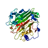
[日本語] English
- PDB-8beg: Structure of Ig-like domains from PrgB -

+
データを開く


IDまたはキーワード:

読み込み中...

-
基本情報

登録情報
データベース: PDB / ID: 8beg
タイトルStructure of Ig-like domains from PrgB
要素PrgB
キーワードCELL ADHESION / Ig-like domians
機能・相同性
機能・相同性情報


extracellular region / metal ion binding / membrane
類似検索 - 分子機能
Antigen I/II, N-terminal / Adhesin P1 N-terminal domain / Glucan-binding protein C/Surface antigen I/II, V-domain / Glucan-binding protein C/Surface antigen I/II, V-domain superfamily / Glucan-binding protein C / Adhesin isopeptide-forming adherence domain / Cell surface antigen, C-terminal / Cell surface antigen C-terminus / Cell surface antigen I/II C2 terminal domain / Immunoglobulin-like - #740 ...Antigen I/II, N-terminal / Adhesin P1 N-terminal domain / Glucan-binding protein C/Surface antigen I/II, V-domain / Glucan-binding protein C/Surface antigen I/II, V-domain superfamily / Glucan-binding protein C / Adhesin isopeptide-forming adherence domain / Cell surface antigen, C-terminal / Cell surface antigen C-terminus / Cell surface antigen I/II C2 terminal domain / Immunoglobulin-like - #740 / KxYKxGKxW signal peptide / KxYKxGKxW signal peptide / LPXTG cell wall anchor motif / Gram-positive cocci surface proteins LPxTG motif profile. / LPXTG cell wall anchor domain / Immunoglobulin-like / Sandwich / Mainly Beta
類似検索 - ドメイン・相同性
生物種Enterococcus faecalis (乳酸球菌)
手法X線回折 / シンクロトロン / 分子置換 / 解像度: 1.84 Å
データ登録者Jarva, M. / Schmitt, A. / Berntsson, R.P.-A.
資金援助 スウェーデン, 2件
組織認可番号
Swedish Research Council2016-03599 スウェーデン
Carl Trygger FoundationCTS 18:39 スウェーデン
引用ジャーナル: Elife / : 2023
タイトル: Structural foundation for the role of enterococcal PrgB in conjugation, biofilm formation, and virulence.
著者: Wei-Sheng Sun / Lena Lassinantti / Michael Järvå / Andreas Schmitt / Josy Ter Beek / Ronnie P-A Berntsson /
要旨: Type 4 Secretion Systems are a main driver for the spread of antibiotic resistance genes and virulence factors in bacteria. In Gram-positives, these secretion systems often rely on surface adhesins ...Type 4 Secretion Systems are a main driver for the spread of antibiotic resistance genes and virulence factors in bacteria. In Gram-positives, these secretion systems often rely on surface adhesins to enhance cellular aggregation and mating-pair formation. One of the best studied adhesins is PrgB from the conjugative plasmid pCF10 of , which has been shown to play major roles in conjugation, biofilm formation, and importantly also in bacterial virulence. Since orthologs exist on a large number of conjugative plasmids in various different species, this makes PrgB a model protein for this widespread virulence factor. After characterizing the polymer adhesin domain of PrgB previously, we here report the structure for almost the entire remainder of PrgB, which reveals that PrgB contains four immunoglobulin (Ig)-like domains. Based on this new insight, we re-evaluate previously studied variants and present new in vivo data where specific domains or conserved residues have been removed. For the first time, we can show a decoupling of cellular aggregation from biofilm formation and conjugation in mutant phenotypes. Based on the presented data, we propose a new functional model to explain how PrgB mediates its different functions. We hypothesize that the Ig-like domains act as a rigid stalk that presents the polymer adhesin domain at the right distance from the cell wall.
履歴
登録2022年10月21日登録サイト: PDBE / 処理サイト: PDBE
改定 1.02022年11月30日Provider: repository / タイプ: Initial release
改定 1.12023年11月1日Group: Data collection / Database references
カテゴリ: chem_comp_atom / chem_comp_bond ...chem_comp_atom / chem_comp_bond / citation / citation_author
Item: _citation.country / _citation.journal_abbrev ..._citation.country / _citation.journal_abbrev / _citation.journal_id_CSD / _citation.journal_id_ISSN / _citation.journal_volume / _citation.pdbx_database_id_DOI / _citation.pdbx_database_id_PubMed / _citation.title / _citation.year / _citation_author.identifier_ORCID / _citation_author.name
改定 1.22024年5月1日Group: Refinement description / カテゴリ: pdbx_initial_refinement_model

-
構造の表示

構造ビューア分子:
MolmilJmol/JSmol

ダウンロードとリンク

-
集合体

登録構造単位
A: PrgB
B: PrgB
ヘテロ分子


分子量 (理論値)分子数
合計 (水以外)143,6914
ポリマ-143,6422
非ポリマー492
14,700816
1


  • 登録構造と同一
  • 登録者・ソフトウェアが定義した集合体
  • 根拠: light scattering, SEC-MALLS and mass phytometry suggests the protein is in a dimer/monomer equilibrium in solution
タイプ名称対称操作
identity operation1_555x,y,z1
Buried area1980 Å2
ΔGint-23 kcal/mol
Surface area56450 Å2
手法PISA
単位格子
Length a, b, c (Å)76.000, 87.500, 224.800
Angle α, β, γ (deg.)90.000, 90.000, 90.000
Int Tables number19
Space group name H-MP212121

-
要素

#1: タンパク質 PrgB


分子量: 71821.109 Da / 分子数: 2 / 由来タイプ: 組換発現 / 詳細: from the conjugative plasmid pCF10 / 由来: (組換発現) Enterococcus faecalis (乳酸球菌) / 遺伝子: prgB / 発現宿主: Escherichia coli BL21(DE3) (大腸菌) / 参照: UniProt: Q04112
#2: 化合物 ChemComp-MG / MAGNESIUM ION / マグネシウムジカチオン


分子量: 24.305 Da / 分子数: 2 / 由来タイプ: 合成 / : Mg
#3: 水 ChemComp-HOH / water


分子量: 18.015 Da / 分子数: 816 / 由来タイプ: 天然 / : H2O
研究の焦点であるリガンドがあるかN

-
実験情報

-
実験

実験手法: X線回折 / 使用した結晶の数: 1

-
試料調製

結晶マシュー密度: 2.6 Å3/Da / 溶媒含有率: 52.73 %
結晶化温度: 293 K / 手法: 蒸気拡散法, シッティングドロップ法 / pH: 7 / 詳細: CaCl2, PEG3350

-
データ収集

回折平均測定温度: 100 K / Serial crystal experiment: N
放射光源由来: シンクロトロン / サイト: ESRF / ビームライン: ID23-1 / 波長: 0.975 Å
検出器タイプ: DECTRIS PILATUS 6M / 検出器: PIXEL / 日付: 2015年12月15日
放射プロトコル: SINGLE WAVELENGTH / 単色(M)・ラウエ(L): M / 散乱光タイプ: x-ray
放射波長波長: 0.975 Å / 相対比: 1
反射解像度: 1.79→47.29 Å / Num. obs: 139448 / % possible obs: 98.6 % / 冗長度: 6.568 % / Biso Wilson estimate: 36.95 Å2 / CC1/2: 0.999 / Rmerge(I) obs: 0.071 / Rrim(I) all: 0.077 / Χ2: 0.812 / Net I/σ(I): 12.43 / Num. measured all: 915892
反射 シェル

Diffraction-ID: 1

解像度 (Å)冗長度 (%)Rmerge(I) obsMean I/σ(I) obsNum. measured obsNum. possibleNum. unique obsCC1/2Rrim(I) all% possible all
1.79-1.96.4341.8390.7713505622692209910.6091.99892.5
1.9-2.036.5080.951.613768221193211550.8771.03399.8
2.03-2.196.9320.5363.0613733819833198110.9530.57999.9
2.19-2.46.590.3274.9712009318264182230.9760.35599.8
2.4-2.696.7980.1749.1811276216611165870.9930.18999.9
2.69-3.16.5740.08718.039640314695146650.9970.09599.8
3.1-3.796.4870.0533.228124212549125230.9990.05499.8
3.79-5.356.140.03743.7660081981497850.9990.0499.7
5.35-47.296.1730.0347.6735235574457080.9990.03299.4

-
解析

ソフトウェア
名称バージョン分類
XSCALEデータスケーリング
PHENIX1.9.2精密化
PDB_EXTRACT3.27データ抽出
XDSデータ削減
PHASER位相決定
精密化構造決定の手法: 分子置換
開始モデル: Alphafold model

解像度: 1.84→47.29 Å / SU ML: 0.29 / 交差検証法: THROUGHOUT / σ(F): 1.33 / 位相誤差: 32.77 / 立体化学のターゲット値: ML
Rfactor反射数%反射
Rfree0.2455 6485 4.98 %
Rwork0.2089 123634 -
obs0.2108 130119 99.63 %
溶媒の処理減衰半径: 0.9 Å / VDWプローブ半径: 1.11 Å / 溶媒モデル: FLAT BULK SOLVENT MODEL
原子変位パラメータBiso max: 100.32 Å2 / Biso mean: 45.0436 Å2 / Biso min: 26.56 Å2
精密化ステップサイクル: final / 解像度: 1.84→47.29 Å
タンパク質核酸リガンド溶媒全体
原子数10052 0 11 816 10879
Biso mean--57.93 47.67 -
残基数----1299
LS精密化 シェル

Refine-ID: X-RAY DIFFRACTION / Rfactor Rfree error: 0 / Total num. of bins used: 30

解像度 (Å)Rfactor RfreeNum. reflection RfreeRfactor RworkNum. reflection RworkNum. reflection all% reflection obs (%)
1.84-1.860.41532260.41024050427699
1.86-1.880.42732080.40134038424699
1.88-1.910.42882220.386441064328100
1.91-1.930.41332340.35344043427799
1.93-1.960.36142290.336140304259100
1.96-1.980.35692070.308640644271100
1.98-2.010.33222170.304441144331100
2.01-2.040.34891930.288640944287100
2.04-2.070.36131780.291841384316100
2.07-2.110.31742120.283140674279100
2.11-2.140.29882190.265541004319100
2.14-2.180.34512050.25154098430399
2.18-2.220.34592250.261440614286100
2.22-2.270.29592140.249140724286100
2.27-2.320.28312170.247841034320100
2.32-2.370.28431960.234841394335100
2.37-2.430.29372200.238640754295100
2.43-2.50.30822010.237641074308100
2.5-2.570.31231950.23941604355100
2.57-2.650.31642060.24841144320100
2.65-2.750.30522180.271841154333100
2.75-2.860.30682310.24841034334100
2.86-2.990.29822030.232741394342100
2.99-3.150.28982360.232941284364100
3.15-3.340.23442190.224141594378100
3.34-3.60.23352240.19641894413100
3.6-3.960.21342240.181941664390100
3.96-4.540.19122310.150841824413100
4.54-5.710.15632310.142542504481100
5.71-47.290.17772440.155844304674100

+
万見について

-
お知らせ

-
2022年2月9日: EMDBエントリの付随情報ファイルのフォーマットが新しくなりました

EMDBエントリの付随情報ファイルのフォーマットが新しくなりました

  • EMDBのヘッダファイルのバージョン3が、公式のフォーマットとなりました。
  • これまでは公式だったバージョン1.9は、アーカイブから削除されます。

関連情報:EMDBヘッダ

外部リンク:wwPDBはEMDBデータモデルのバージョン3へ移行します

-
2020年8月12日: 新型コロナ情報

新型コロナ情報

URL: https://pdbj.org/emnavi/covid19.php

新ページ: EM Navigatorに新型コロナウイルスの特設ページを開設しました。

関連情報:Covid-19情報 / 2020年3月5日: 新型コロナウイルスの構造データ

+
2020年3月5日: 新型コロナウイルスの構造データ

新型コロナウイルスの構造データ

関連情報:万見生物種 / 2020年8月12日: 新型コロナ情報

外部リンク:COVID-19特集ページ - PDBj / 今月の分子2020年2月:コロナウイルスプロテーアーゼ

+
2019年1月31日: EMDBのIDの桁数の変更

EMDBのIDの桁数の変更

  • EMDBエントリに付与されているアクセスコード(EMDB-ID)は4桁の数字(例、EMD-1234)でしたが、間もなく枯渇します。これまでの4桁のID番号は4桁のまま変更されませんが、4桁の数字を使い切った後に発行されるIDは5桁以上の数字(例、EMD-12345)になります。5桁のIDは2019年の春頃から発行される見通しです。
  • EM Navigator/万見では、接頭語「EMD-」は省略されています。

関連情報:Q: 「EMD」とは何ですか? / 万見/EM NavigatorにおけるID/アクセスコードの表記

外部リンク:EMDB Accession Codes are Changing Soon! / PDBjへお問い合わせ

+
2017年7月12日: PDB大規模アップデート

PDB大規模アップデート

  • 新バージョンのPDBx/mmCIF辞書形式に基づくデータがリリースされました。
  • 今回の更新はバージョン番号が4から5になる大規模なもので、全エントリデータの書き換えが行われる「Remediation」というアップデートに該当します。
  • このバージョンアップで、電子顕微鏡の実験手法に関する多くの項目の書式が改定されました(例:em_softwareなど)。
  • EM NavigatorとYorodumiでも、この改定に基づいた表示内容になります。

外部リンク:wwPDB Remediation / OneDepデータ基準に準拠した、より強化された内容のモデル構造ファイルが、PDBアーカイブで公開されました。

-
万見 (Yorodumi)

幾万の構造データを、幾万の視点から

  • 万見(Yorodumi)は、EMDB/PDB/SASBDBなどの構造データを閲覧するためのページです。
  • EM Navigatorの詳細ページの後継、Omokage検索のフロントエンドも兼ねています。

関連情報:EMDB / PDB / SASBDB / 3つのデータバンクの比較 / 万見検索 / 2016年8月31日: 新しいEM Navigatorと万見 / 万見文献 / Jmol/JSmol / 機能・相同性情報 / 新しいEM Navigatorと万見の変更点

他の情報も見る