 Movie
Movie Controller

[English] 日本語
 Yorodumi
- PDB-8bcs: X-ray crystal structure of a de novo designed helix-loop-helix ho... -
+ Open data

- Basic information
| Entry | Database: PDB / ID: 8bcs | ||||||||||||
|---|---|---|---|---|---|---|---|---|---|---|---|---|---|
| Title | X-ray crystal structure of a de novo designed helix-loop-helix homodimer in an anti arrangement, CC-HP1.0 | ||||||||||||
 Components Components | CC-HP1.0 | ||||||||||||
 Keywords Keywords | DE NOVO PROTEIN / coiled coil / 4-helix bundle / de novo protein design / helix-loop-helix dimer | ||||||||||||
| Function / homology | ACETATE ION Function and homology information Function and homology information | ||||||||||||
| Biological species | synthetic construct (others) | ||||||||||||
| Method |  X-RAY DIFFRACTION / SYNCHROTRON / MOLECULAR REPLACEMENT / Resolution: 2.1 Å X-RAY DIFFRACTION / SYNCHROTRON / MOLECULAR REPLACEMENT / Resolution: 2.1 Å | ||||||||||||
 Authors Authors | Edgell, C.L. / Mylemans, B. / Naudin, E.A. / Smith, A.J. / Savery, N.J. / Woolfson, D.N. | ||||||||||||
| Funding support |  United Kingdom, 3items United Kingdom, 3items
| ||||||||||||
 Citation Citation | Journal: Acs Synth Biol / Year: 2023 Title: Design and Selection of Heterodimerizing Helical Hairpins for Synthetic Biology. Authors: Smith, A.J. / Naudin, E.A. / Edgell, C.L. / Baker, E.G. / Mylemans, B. / FitzPatrick, L. / Herman, A. / Rice, H.M. / Andrews, D.M. / Tigue, N. / Woolfson, D.N. / Savery, N.J. | ||||||||||||
| History |
|
- Structure visualization
| Structure viewer | Molecule: MolmilJmol/JSmol |
|---|
- Downloads & links
-Download
| PDBx/mmCIF format | 8bcs.cif.gz | 31.2 KB | Display | PDBx/mmCIF format |
|---|---|---|---|---|
| PDB format | pdb8bcs.ent.gz | 18.7 KB | Display | PDB format |
| PDBx/mmJSON format | 8bcs.json.gz | Tree view | PDBx/mmJSON format | |
| Others |  Other downloads Other downloads |
-Validation report
| Arichive directory | https://data.pdbj.org/pub/pdb/validation_reports/bc/8bcsftp://data.pdbj.org/pub/pdb/validation_reports/bc/8bcs | HTTPS FTP |
|---|
-Related structure data
| Related structure data |  8bctC 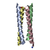 6q5sS S: Starting model for refinement C: citing same article ( |
|---|---|
| Similar structure data |
-Links
 PDBj
PDBj
- Assembly
| Deposited unit | 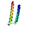
| ||||||||||||
|---|---|---|---|---|---|---|---|---|---|---|---|---|---|
| 1 | 
| ||||||||||||
| Unit cell |
|
-Components
| #1: Protein | Mass: 5275.271 Da / Num. of mol.: 1 / Source method: obtained synthetically / Source: (synth.) synthetic construct (others) |
|---|---|
| #2: Chemical | ChemComp-ACT /   Mass: 59.044 Da / Num. of mol.: 1 / Source method: obtained synthetically / Formula: C2H3O2 Mass: 59.044 Da / Num. of mol.: 1 / Source method: obtained synthetically / Formula: C2H3O2 |
| Has ligand of interest | N |
| Has protein modification | N |
-Experimental details
-Experiment
| Experiment | Method: X-RAY DIFFRACTION / Number of used crystals: 1 |
|---|
- Sample preparation
| Crystal | Density Matthews: 2.16 Å3/Da / Density % sol: 42.93 % |
|---|---|
| Crystal grow | Temperature: 293 K / Method: vapor diffusion, sitting drop / pH: 6.5 Details: 0.2 M sodium acetate trihydrate, 0.1 M sodium cacodylate, 30% w/v PEG 8000, pH 6.5 |
-Data collection
| Diffraction | Mean temperature: 100 K / Serial crystal experiment: N | |||||||||||||||||||||
|---|---|---|---|---|---|---|---|---|---|---|---|---|---|---|---|---|---|---|---|---|---|---|
| Diffraction source | Source: SYNCHROTRON / Site: Diamond / Beamline: I04 / Wavelength: 0.9795 Å | |||||||||||||||||||||
| Detector | Type: DECTRIS EIGER2 XE 16M / Detector: PIXEL / Date: Oct 12, 2019 | |||||||||||||||||||||
| Radiation | Protocol: SINGLE WAVELENGTH / Monochromatic (M) / Laue (L): M / Scattering type: x-ray | |||||||||||||||||||||
| Radiation wavelength | Wavelength: 0.9795 Å / Relative weight: 1 | |||||||||||||||||||||
| Reflection | Resolution: 1.99→31.56 Å / Num. obs: 3400 / % possible obs: 100 % / Redundancy: 16.5 % / Biso Wilson estimate: 53.42 Å2 / CC1/2: 1 / Rrim(I) all: 0.038 / Net I/σ(I): 27 | |||||||||||||||||||||
| Reflection shell | Diffraction-ID: 1
|
- Processing
| Software |
| ||||||||||||||||||||||||||||||||||||||||||
|---|---|---|---|---|---|---|---|---|---|---|---|---|---|---|---|---|---|---|---|---|---|---|---|---|---|---|---|---|---|---|---|---|---|---|---|---|---|---|---|---|---|---|---|
| Refinement | Method to determine structure: MOLECULAR REPLACEMENT Starting model: 6Q5S Resolution: 2.1→31.56 Å / SU ML: 0.2354 / Cross valid method: FREE R-VALUE / σ(F): 1.38 / Phase error: 26.8234 Stereochemistry target values: GeoStd + Monomer Library + CDL v1.2
| ||||||||||||||||||||||||||||||||||||||||||
| Solvent computation | Shrinkage radii: 0.9 Å / VDW probe radii: 1.11 Å / Solvent model: FLAT BULK SOLVENT MODEL | ||||||||||||||||||||||||||||||||||||||||||
| Displacement parameters | Biso mean: 67.81 Å2 | ||||||||||||||||||||||||||||||||||||||||||
| Refinement step | Cycle: LAST / Resolution: 2.1→31.56 Å
| ||||||||||||||||||||||||||||||||||||||||||
| Refine LS restraints |
| ||||||||||||||||||||||||||||||||||||||||||
| LS refinement shell |
| ||||||||||||||||||||||||||||||||||||||||||
| Refinement TLS params. | Method: refined / Origin x: -3.23793364477 Å / Origin y: -7.9972962818 Å / Origin z: 4.18976254378 Å
| ||||||||||||||||||||||||||||||||||||||||||
| Refinement TLS group | Selection details: chain 'A' and (resid 1 through 49 ) |
