 Movie
Movie Controller


+ Open data

- Basic information
| Entry | Database: PDB / ID: 8b8d | |||||||||
|---|---|---|---|---|---|---|---|---|---|---|
| Title | multimerization domain of Gaboon Viper Virus 1 | |||||||||
 Components Components | Phosphoprotein | |||||||||
 Keywords Keywords | VIRAL PROTEIN / phosphoprotein / RNA polymerase cofactor | |||||||||
| Function / homology | Borna disease virus P24 / Borna disease virus P24 protein / Phosphoprotein Function and homology information Function and homology information | |||||||||
| Biological species |  Gaboon viper virus 1 Gaboon viper virus 1 | |||||||||
| Method |  X-RAY DIFFRACTION / SYNCHROTRON / SAD / Resolution: 2.4 Å X-RAY DIFFRACTION / SYNCHROTRON / SAD / Resolution: 2.4 Å | |||||||||
 Authors Authors | Tarbouriech, N. / Legrand, P. / Bouhris, J.M. / Horie, M. / Tomonaga, K. / Crepin, T. | |||||||||
| Funding support |  France, France,  Japan, 2items Japan, 2items
| |||||||||
 Citation Citation | Journal: Viruses / Year: 2022 Title: Borna Disease Virus 1 Phosphoprotein Forms a Tetramer and Interacts with Host Factors Involved in DNA Double-Strand Break Repair and mRNA Processing. Authors: Tarbouriech, N. / Chenavier, F. / Kawasaki, J. / Bachiri, K. / Bourhis, J.M. / Legrand, P. / Freslon, L.L. / Laurent, E.M.N. / Suberbielle, E. / Ruigrok, R.W.H. / Tomonaga, K. / Gonzalez- ...Authors: Tarbouriech, N. / Chenavier, F. / Kawasaki, J. / Bachiri, K. / Bourhis, J.M. / Legrand, P. / Freslon, L.L. / Laurent, E.M.N. / Suberbielle, E. / Ruigrok, R.W.H. / Tomonaga, K. / Gonzalez-Dunia, D. / Horie, M. / Coyaud, E. / Crepin, T. | |||||||||
| History |
|
- Structure visualization
| Structure viewer | Molecule: MolmilJmol/JSmol |
|---|
- Downloads & links
-Download
| PDBx/mmCIF format | 8b8d.cif.gz | 94.5 KB | Display | PDBx/mmCIF format |
|---|---|---|---|---|
| PDB format | pdb8b8d.ent.gz | 72.9 KB | Display | PDB format |
| PDBx/mmJSON format | 8b8d.json.gz | Tree view | PDBx/mmJSON format | |
| Others |  Other downloads Other downloads |
-Validation report
| Arichive directory | https://data.pdbj.org/pub/pdb/validation_reports/b8/8b8dftp://data.pdbj.org/pub/pdb/validation_reports/b8/8b8d | HTTPS FTP |
|---|
-Related structure data
| Related structure data | 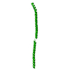 8b8aC  8b8bC C: citing same article ( |
|---|---|
| Similar structure data | |
| Other databases |
|
-Links
 PDBj
PDBj- Assembly
| Deposited unit | 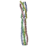
| ||||||||
|---|---|---|---|---|---|---|---|---|---|
| 1 |
| ||||||||
| Unit cell |
|
-Components
| #1: Protein | Mass: 12920.860 Da / Num. of mol.: 4 Source method: isolated from a genetically manipulated source Source: (gene. exp.) Gaboon viper virus 1 / Plasmid: pETM11 / Production host:  #2: Water | ChemComp-HOH / |  Mass: 18.015 Da / Num. of mol.: 149 / Source method: isolated from a natural source / Formula: H2O Mass: 18.015 Da / Num. of mol.: 149 / Source method: isolated from a natural source / Formula: H2O |
|---|
-Experimental details
-Experiment
| Experiment | Method: X-RAY DIFFRACTION / Number of used crystals: 1 |
|---|
- Sample preparation
| Crystal | Density Matthews: 2.91 Å3/Da / Density % sol: 57.7 % |
|---|---|
| Crystal grow | Temperature: 293 K / Method: vapor diffusion, hanging drop / pH: 7.5 Details: 100 mM HEPES pH 7.5, 18-22 % PEG 3350, 200 mM NaSCN |
-Data collection
| Diffraction |
| ||||||||||||||||||||||||
|---|---|---|---|---|---|---|---|---|---|---|---|---|---|---|---|---|---|---|---|---|---|---|---|---|---|
| Diffraction source |
| ||||||||||||||||||||||||
| Detector |
| ||||||||||||||||||||||||
| Radiation |
| ||||||||||||||||||||||||
| Radiation wavelength |
| ||||||||||||||||||||||||
| Reflection | Entry-ID: 8B8D / CC1/2: 0.999
| ||||||||||||||||||||||||
| Reflection shell |
|
- Processing
| Software |
| ||||||||||||||||||||||||||||||||||||||||||||||||||||||||||||
|---|---|---|---|---|---|---|---|---|---|---|---|---|---|---|---|---|---|---|---|---|---|---|---|---|---|---|---|---|---|---|---|---|---|---|---|---|---|---|---|---|---|---|---|---|---|---|---|---|---|---|---|---|---|---|---|---|---|---|---|---|---|
| Refinement | Method to determine structure: SAD / Resolution: 2.4→48.71 Å / Cor.coef. Fo:Fc: 0.941 / Cor.coef. Fo:Fc free: 0.908 / SU B: 10.234 / SU ML: 0.238 / Cross valid method: THROUGHOUT / σ(F): 0 / ESU R: 0.505 / ESU R Free: 0.352 / Stereochemistry target values: MAXIMUM LIKELIHOOD Details: HYDROGENS HAVE BEEN ADDED IN THE RIDING POSITIONS U VALUES : REFINED INDIVIDUALLY
| ||||||||||||||||||||||||||||||||||||||||||||||||||||||||||||
| Solvent computation | Ion probe radii: 0.8 Å / Shrinkage radii: 0.8 Å / VDW probe radii: 1.2 Å / Solvent model: MASK | ||||||||||||||||||||||||||||||||||||||||||||||||||||||||||||
| Displacement parameters | Biso max: 131.63 Å2 / Biso mean: 52.184 Å2 / Biso min: 10.49 Å2
| ||||||||||||||||||||||||||||||||||||||||||||||||||||||||||||
| Refinement step | Cycle: final / Resolution: 2.4→48.71 Å
| ||||||||||||||||||||||||||||||||||||||||||||||||||||||||||||
| Refine LS restraints |
| ||||||||||||||||||||||||||||||||||||||||||||||||||||||||||||
| LS refinement shell | Resolution: 2.4→2.463 Å / Rfactor Rfree error: 0 / Total num. of bins used: 20
|
