 Movie
Movie Controller


+ Open data

- Basic information
| Entry | Database: PDB / ID: 8b4g | ||||||||||||
|---|---|---|---|---|---|---|---|---|---|---|---|---|---|
| Title | Structure of a fungal LPMO bound to ligands | ||||||||||||
 Components Components | Gh61 isozyme a | ||||||||||||
 Keywords Keywords | OXIDOREDUCTASE / Monooxygenase / dihydroxyacetone / Lytic Polysaccharide Monooxygenase / Thermoascus aurantiacus | ||||||||||||
| Function / homology |  Function and homology information Function and homology informationlytic cellulose monooxygenase (C4-dehydrogenating) / cellulose catabolic process / monooxygenase activity / extracellular region / metal ion binding Similarity search - Function | ||||||||||||
| Biological species |  Thermoascus aurantiacus (fungus) Thermoascus aurantiacus (fungus) | ||||||||||||
| Method |  X-RAY DIFFRACTION / SYNCHROTRON / MOLECULAR REPLACEMENT / Resolution: 1.496 Å X-RAY DIFFRACTION / SYNCHROTRON / MOLECULAR REPLACEMENT / Resolution: 1.496 Å | ||||||||||||
 Authors Authors | Banerjee, S. / Huang, Z. / Brander, S. / Johansen, K.S. / Lo Leggio, L. | ||||||||||||
| Funding support |  Denmark, Denmark,  Sweden, 3items Sweden, 3items
| ||||||||||||
 Citation Citation | Journal: To Be Published Title: Structure of a fungal LPMO bound to ligands Authors: Banerjee, S. / Huang, Z. / Brander, S. / Johansen, K.S. / Lo Leggio, L. | ||||||||||||
| History |
|
- Structure visualization
| Structure viewer | Molecule: MolmilJmol/JSmol |
|---|
- Downloads & links
-Download
| PDBx/mmCIF format | 8b4g.cif.gz | 72.4 KB | Display | PDBx/mmCIF format |
|---|---|---|---|---|
| PDB format | pdb8b4g.ent.gz | Display | PDB format | |
| PDBx/mmJSON format | 8b4g.json.gz | Tree view | PDBx/mmJSON format | |
| Others |  Other downloads Other downloads |
-Validation report
| Arichive directory | https://data.pdbj.org/pub/pdb/validation_reports/b4/8b4gftp://data.pdbj.org/pub/pdb/validation_reports/b4/8b4g | HTTPS FTP |
|---|
-Related structure data
| Related structure data | 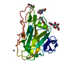 3zudS S: Starting model for refinement |
|---|---|
| Similar structure data |
-Links
 PDBj
PDBj- Assembly
| Deposited unit | 
| ||||||||
|---|---|---|---|---|---|---|---|---|---|
| 1 |
| ||||||||
| Unit cell |
|
-Components
-Protein , 1 types, 1 molecules AAA
| #1: Protein | Mass: 24418.043 Da / Num. of mol.: 1 Source method: isolated from a genetically manipulated source Source: (gene. exp.) Thermoascus aurantiacus (fungus) / Production host: |
|---|
-Sugars , 2 types, 2 molecules 
| #3: Sugar | ChemComp-NAG /  Type: D-saccharide, beta linking / Mass: 221.208 Da / Num. of mol.: 1 / Source method: obtained synthetically / Formula: C8H15NO6 Type: D-saccharide, beta linking / Mass: 221.208 Da / Num. of mol.: 1 / Source method: obtained synthetically / Formula: C8H15NO6 |
|---|---|
| #5: Sugar | ChemComp-2HA /  Type: saccharide / Mass: 90.078 Da / Num. of mol.: 1 / Source method: obtained synthetically / Formula: C3H6O3 / Feature type: SUBJECT OF INVESTIGATION Type: saccharide / Mass: 90.078 Da / Num. of mol.: 1 / Source method: obtained synthetically / Formula: C3H6O3 / Feature type: SUBJECT OF INVESTIGATION |
-Non-polymers , 4 types, 280 molecules 


| #2: Chemical | ChemComp-CU /  Mass: 63.546 Da / Num. of mol.: 1 / Source method: obtained synthetically / Formula: Cu Mass: 63.546 Da / Num. of mol.: 1 / Source method: obtained synthetically / Formula: Cu | ||||
|---|---|---|---|---|---|
| #4: Chemical |  Mass: 72.063 Da / Num. of mol.: 2 / Source method: obtained synthetically / Formula: C3H4O2 Mass: 72.063 Da / Num. of mol.: 2 / Source method: obtained synthetically / Formula: C3H4O2#6: Chemical |  Mass: 35.453 Da / Num. of mol.: 2 / Source method: obtained synthetically / Formula: Cl Mass: 35.453 Da / Num. of mol.: 2 / Source method: obtained synthetically / Formula: Cl#7: Water | ChemComp-HOH / | Mass: 18.015 Da / Num. of mol.: 275 / Source method: isolated from a natural source / Formula: H2O |
-Details
| Has ligand of interest | Y |
|---|---|
| Has protein modification | Y |
-Experimental details
-Experiment
| Experiment | Method: X-RAY DIFFRACTION / Number of used crystals: 1 |
|---|
- Sample preparation
| Crystal | Density Matthews: 2.23 Å3/Da / Density % sol: 44.81 % |
|---|---|
| Crystal grow | Temperature: 293 K / Method: vapor diffusion, sitting drop Details: 0.1 M HEPES pH 7.5, 20m M MgCl2 and 22 %(w/v) polyacrylic acid 5100 sodium salt |
-Data collection
| Diffraction | Mean temperature: 100 K / Serial crystal experiment: N |
|---|---|
| Diffraction source | Source: SYNCHROTRON / Site: ESRF  / Beamline: ID23-2 / Wavelength: 0.8731 Å / Beamline: ID23-2 / Wavelength: 0.8731 Å |
| Detector | Type: DECTRIS PILATUS3 X 2M / Detector: PIXEL / Date: Jun 4, 2021 |
| Radiation | Protocol: SINGLE WAVELENGTH / Monochromatic (M) / Laue (L): M / Scattering type: x-ray |
| Radiation wavelength | Wavelength: 0.8731 Å / Relative weight: 1 |
| Reflection | Resolution: 1.496→87.31 Å / Num. obs: 34235 / % possible obs: 99.3 % / Redundancy: 6.6 % / CC1/2: 0.996 / Net I/σ(I): 7.9 |
| Reflection shell | Resolution: 1.5→1.52 Å / Num. unique obs: 1426 / CC1/2: 0.509 |
- Processing
| Software |
| |||||||||||||||||||||||||||||||||||||||||||||||||||||||||||||||||||||||||||||||||||||||||||||||||||||||||||||||||||||||||||||||||||||||||||||||||||||||||||||||||||||||||||||||||||||||||||||||||||||||||||||||||||||||||||||||||||||||
|---|---|---|---|---|---|---|---|---|---|---|---|---|---|---|---|---|---|---|---|---|---|---|---|---|---|---|---|---|---|---|---|---|---|---|---|---|---|---|---|---|---|---|---|---|---|---|---|---|---|---|---|---|---|---|---|---|---|---|---|---|---|---|---|---|---|---|---|---|---|---|---|---|---|---|---|---|---|---|---|---|---|---|---|---|---|---|---|---|---|---|---|---|---|---|---|---|---|---|---|---|---|---|---|---|---|---|---|---|---|---|---|---|---|---|---|---|---|---|---|---|---|---|---|---|---|---|---|---|---|---|---|---|---|---|---|---|---|---|---|---|---|---|---|---|---|---|---|---|---|---|---|---|---|---|---|---|---|---|---|---|---|---|---|---|---|---|---|---|---|---|---|---|---|---|---|---|---|---|---|---|---|---|---|---|---|---|---|---|---|---|---|---|---|---|---|---|---|---|---|---|---|---|---|---|---|---|---|---|---|---|---|---|---|---|---|---|---|---|---|---|---|---|---|---|---|---|---|---|---|---|---|---|
| Refinement | Method to determine structure: MOLECULAR REPLACEMENT Starting model: 3ZUD Resolution: 1.496→43.656 Å / Cor.coef. Fo:Fc: 0.976 / Cor.coef. Fo:Fc free: 0.964 / WRfactor Rfree: 0.168 / WRfactor Rwork: 0.138 / SU B: 1.575 / SU ML: 0.055 / Average fsc free: 0.9201 / Average fsc work: 0.927 / Cross valid method: FREE R-VALUE / ESU R: 0.068 / ESU R Free: 0.071 Details: Hydrogens have been added in their riding positions
| |||||||||||||||||||||||||||||||||||||||||||||||||||||||||||||||||||||||||||||||||||||||||||||||||||||||||||||||||||||||||||||||||||||||||||||||||||||||||||||||||||||||||||||||||||||||||||||||||||||||||||||||||||||||||||||||||||||||
| Solvent computation | Ion probe radii: 0.8 Å / Shrinkage radii: 0.8 Å / VDW probe radii: 1.2 Å / Solvent model: MASK BULK SOLVENT | |||||||||||||||||||||||||||||||||||||||||||||||||||||||||||||||||||||||||||||||||||||||||||||||||||||||||||||||||||||||||||||||||||||||||||||||||||||||||||||||||||||||||||||||||||||||||||||||||||||||||||||||||||||||||||||||||||||||
| Displacement parameters | Biso mean: 14.843 Å2
| |||||||||||||||||||||||||||||||||||||||||||||||||||||||||||||||||||||||||||||||||||||||||||||||||||||||||||||||||||||||||||||||||||||||||||||||||||||||||||||||||||||||||||||||||||||||||||||||||||||||||||||||||||||||||||||||||||||||
| Refinement step | Cycle: LAST / Resolution: 1.496→43.656 Å
| |||||||||||||||||||||||||||||||||||||||||||||||||||||||||||||||||||||||||||||||||||||||||||||||||||||||||||||||||||||||||||||||||||||||||||||||||||||||||||||||||||||||||||||||||||||||||||||||||||||||||||||||||||||||||||||||||||||||
| Refine LS restraints |
| |||||||||||||||||||||||||||||||||||||||||||||||||||||||||||||||||||||||||||||||||||||||||||||||||||||||||||||||||||||||||||||||||||||||||||||||||||||||||||||||||||||||||||||||||||||||||||||||||||||||||||||||||||||||||||||||||||||||
| LS refinement shell | Refine-ID: X-RAY DIFFRACTION / Total num. of bins used: 20
|
