 Movie
Movie Controller


+ Open data

- Basic information
| Entry | Database: PDB / ID: 8b1p | ||||||
|---|---|---|---|---|---|---|---|
| Title | Crystal structure of SUDV VP40 CCS mutant | ||||||
 Components Components | Matrix protein VP40 | ||||||
 Keywords Keywords | VIRAL PROTEIN / Ebola virus / SUDV / VP40 / matrix protein / dimer | ||||||
| Function / homology |  Function and homology information Function and homology informationhost cell endomembrane system / host cell late endosome membrane / viral budding via host ESCRT complex / structural constituent of virion / symbiont-mediated suppression of host innate immune response / ribonucleoprotein complex / host cell plasma membrane / virion membrane / RNA binding / membrane Similarity search - Function | ||||||
| Biological species |  Sudan ebolavirus Sudan ebolavirus | ||||||
| Method |  X-RAY DIFFRACTION / SYNCHROTRON / MOLECULAR REPLACEMENT / Resolution: 1.7 Å X-RAY DIFFRACTION / SYNCHROTRON / MOLECULAR REPLACEMENT / Resolution: 1.7 Å | ||||||
 Authors Authors | Werner, A.-D. / Becker, S. | ||||||
| Funding support |  Germany, 1items Germany, 1items
| ||||||
 Citation Citation | Journal: Structure / Year: 2023 Title: The C-terminus of Sudan ebolavirus VP40 contains a functionally important CX n C motif, a target for redox modifications. Authors: Werner, A.D. / Schauflinger, M. / Norris, M.J. / Kluver, M. / Trodler, A. / Herwig, A. / Brandstadter, C. / Dillenberger, M. / Klebe, G. / Heine, A. / Saphire, E.O. / Becker, K. / Becker, S. | ||||||
| History |
|
- Structure visualization
| Structure viewer | Molecule: MolmilJmol/JSmol |
|---|
- Downloads & links
-Download
| PDBx/mmCIF format | 8b1p.cif.gz | 129.2 KB | Display | PDBx/mmCIF format |
|---|---|---|---|---|
| PDB format | pdb8b1p.ent.gz | 82.7 KB | Display | PDB format |
| PDBx/mmJSON format | 8b1p.json.gz | Tree view | PDBx/mmJSON format | |
| Others |  Other downloads Other downloads |
-Validation report
| Arichive directory | https://data.pdbj.org/pub/pdb/validation_reports/b1/8b1pftp://data.pdbj.org/pub/pdb/validation_reports/b1/8b1p | HTTPS FTP |
|---|
-Related structure data
| Related structure data |  8b1oC 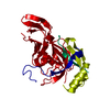 8b3xC  4ld8S S: Starting model for refinement C: citing same article ( |
|---|---|
| Similar structure data |
-Links
 PDBj
PDBj
- Assembly
| Deposited unit | 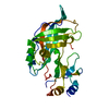
| ||||||||||||
|---|---|---|---|---|---|---|---|---|---|---|---|---|---|
| 1 | 
| ||||||||||||
| Unit cell |
|
-Components
| #1: Protein | Mass: 32571.492 Da / Num. of mol.: 1 Source method: isolated from a genetically manipulated source Source: (gene. exp.) Sudan ebolavirus / Strain: Human/Uganda/Gulu/2000 / Gene: VP40 / Production host:  |
|---|---|
| #2: Water | ChemComp-HOH /  Mass: 18.015 Da / Num. of mol.: 146 / Source method: isolated from a natural source / Formula: H2O Mass: 18.015 Da / Num. of mol.: 146 / Source method: isolated from a natural source / Formula: H2O |
-Experimental details
-Experiment
| Experiment | Method: X-RAY DIFFRACTION / Number of used crystals: 1 |
|---|
- Sample preparation
| Crystal | Density Matthews: 2.15 Å3/Da / Density % sol: 42.71 % |
|---|---|
| Crystal grow | Temperature: 293 K / Method: vapor diffusion, sitting drop Details: Morpheus A7 30% (w/v) GOL_4P4K (glycerol and PEG4000) 0.1 M morpheus buffer 2 (HEPES and MOPS) 60 mM morpheus divalents (MgCl2, CaCl2) |
-Data collection
| Diffraction | Mean temperature: 100 K / Serial crystal experiment: N |
|---|---|
| Diffraction source | Source: SYNCHROTRON / Site: SLS  / Beamline: X06SA / Wavelength: 0.999998 Å / Beamline: X06SA / Wavelength: 0.999998 Å |
| Detector | Type: DECTRIS EIGER X 16M / Detector: PIXEL / Date: Apr 27, 2021 |
| Radiation | Protocol: SINGLE WAVELENGTH / Monochromatic (M) / Laue (L): M / Scattering type: x-ray |
| Radiation wavelength | Wavelength: 0.999998 Å / Relative weight: 1 |
| Reflection | Resolution: 1.6→48.11 Å / Num. obs: 35310 / % possible obs: 98.4 % / Redundancy: 3.8 % / Biso Wilson estimate: 26.9 Å2 / CC1/2: 0.999 / Net I/σ(I): 19 |
| Reflection shell | Resolution: 1.6→1.63 Å / Redundancy: 3.8 % / Mean I/σ(I) obs: 3.5 / Num. unique obs: 1709 / CC1/2: 0.972 / % possible all: 95.8 |
- Processing
| Software |
| ||||||||||||||||||||||||||||||||||||||||||||||||||||||||||||||||||||||||||||||||||||||||||||||||||||
|---|---|---|---|---|---|---|---|---|---|---|---|---|---|---|---|---|---|---|---|---|---|---|---|---|---|---|---|---|---|---|---|---|---|---|---|---|---|---|---|---|---|---|---|---|---|---|---|---|---|---|---|---|---|---|---|---|---|---|---|---|---|---|---|---|---|---|---|---|---|---|---|---|---|---|---|---|---|---|---|---|---|---|---|---|---|---|---|---|---|---|---|---|---|---|---|---|---|---|---|---|---|
| Refinement | Method to determine structure: MOLECULAR REPLACEMENT Starting model: 4LD8 Resolution: 1.7→48.11 Å / SU ML: 0.1767 / Cross valid method: FREE R-VALUE / σ(F): 1.36 / Phase error: 25.0814 Stereochemistry target values: GeoStd + Monomer Library + CDL v1.2
| ||||||||||||||||||||||||||||||||||||||||||||||||||||||||||||||||||||||||||||||||||||||||||||||||||||
| Solvent computation | Shrinkage radii: 0.9 Å / VDW probe radii: 1.11 Å / Solvent model: FLAT BULK SOLVENT MODEL | ||||||||||||||||||||||||||||||||||||||||||||||||||||||||||||||||||||||||||||||||||||||||||||||||||||
| Displacement parameters | Biso mean: 41.94 Å2 | ||||||||||||||||||||||||||||||||||||||||||||||||||||||||||||||||||||||||||||||||||||||||||||||||||||
| Refinement step | Cycle: LAST / Resolution: 1.7→48.11 Å
| ||||||||||||||||||||||||||||||||||||||||||||||||||||||||||||||||||||||||||||||||||||||||||||||||||||
| Refine LS restraints |
| ||||||||||||||||||||||||||||||||||||||||||||||||||||||||||||||||||||||||||||||||||||||||||||||||||||
| LS refinement shell |
| ||||||||||||||||||||||||||||||||||||||||||||||||||||||||||||||||||||||||||||||||||||||||||||||||||||
| Refinement TLS params. | Method: refined / Refine-ID: X-RAY DIFFRACTION
| ||||||||||||||||||||||||||||||||||||||||||||||||||||||||||||||||||||||||||||||||||||||||||||||||||||
| Refinement TLS group |
|
