 Movie
Movie Controller

[English] 日本語
 Yorodumi
- PDB-8b1n: Crystal structure of TrmD-Tm1570 from Calditerrivibrio nitroreduc... -
+ Open data

- Basic information
| Entry | Database: PDB / ID: 8b1n | |||||||||
|---|---|---|---|---|---|---|---|---|---|---|
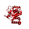
| Title | Crystal structure of TrmD-Tm1570 from Calditerrivibrio nitroreducens in complex with S-adenosyl-L-methionine | |||||||||
 Components Components | tRNA (guanine-N(1)-)-methyltransferase | |||||||||
 Keywords Keywords | TRANSFERASE / TrmD-Tm1570 / TrmD / Tm1570 / Methyl Transferase / Knotted Protein / Double Knotted Protein | |||||||||
| Function / homology |  Function and homology information Function and homology informationtRNA (guanine37-N1)-methyltransferase / tRNA (guanine(37)-N1)-methyltransferase activity / tRNA N1-guanine methylation / metal ion binding / cytosol Similarity search - Function | |||||||||
| Biological species |  Calditerrivibrio nitroreducens DSM 19672 (bacteria) Calditerrivibrio nitroreducens DSM 19672 (bacteria) | |||||||||
| Method |  X-RAY DIFFRACTION / SYNCHROTRON / MOLECULAR REPLACEMENT / Resolution: 2 Å X-RAY DIFFRACTION / SYNCHROTRON / MOLECULAR REPLACEMENT / Resolution: 2 Å | |||||||||
 Authors Authors | Kluza, A. / Lewandowska, I. / Augustyniak, R. / Sulkowska, J. | |||||||||
| Funding support |  Poland, European Union, 2items Poland, European Union, 2items
| |||||||||
 Citation Citation | Journal: Front Mol Biosci / Year: 2023 Title: Are there double knots in proteins? Prediction and in vitro verification based on TrmD-Tm1570 fusion from C. nitroreducens. Authors: Perlinska, A.P. / Nguyen, M.L. / Pilla, S.P. / Staszor, E. / Lewandowska, I. / Bernat, A. / Purta, E. / Augustyniak, R. / Bujnicki, J.M. / Sulkowska, J.I. | |||||||||
| History |
|
- Structure visualization
| Structure viewer | Molecule: MolmilJmol/JSmol |
|---|
- Downloads & links
-Download
| PDBx/mmCIF format | 8b1n.cif.gz | 413.8 KB | Display | PDBx/mmCIF format |
|---|---|---|---|---|
| PDB format | pdb8b1n.ent.gz | 300.3 KB | Display | PDB format |
| PDBx/mmJSON format | 8b1n.json.gz | Tree view | PDBx/mmJSON format | |
| Others |  Other downloads Other downloads |
-Validation report
| Arichive directory | https://data.pdbj.org/pub/pdb/validation_reports/b1/8b1nftp://data.pdbj.org/pub/pdb/validation_reports/b1/8b1n | HTTPS FTP |
|---|
-Related structure data

-Links
 PDBj
PDBj
- Assembly
| Deposited unit | 
| ||||||||||||
|---|---|---|---|---|---|---|---|---|---|---|---|---|---|
| 1 |
| ||||||||||||
| Unit cell |
| ||||||||||||
| Components on special symmetry positions |
|
-Components
-Protein , 1 types, 2 molecules AB
| #1: Protein | Mass: 49997.066 Da / Num. of mol.: 2 Source method: isolated from a genetically manipulated source Source: (gene. exp.) Calditerrivibrio nitroreducens DSM 19672 (bacteria)Strain: DSM 19672 / NBRC 101217 / Yu37-1 / Gene: trmD, Calni_2012 / Production host: References: UniProt: E4THH1, tRNA (guanine37-N1)-methyltransferase |
|---|
-Non-polymers , 5 types, 683 molecules 


| #2: Chemical |  Mass: 24.305 Da / Num. of mol.: 2 / Source method: obtained synthetically / Formula: Mg / Feature type: SUBJECT OF INVESTIGATION Mass: 24.305 Da / Num. of mol.: 2 / Source method: obtained synthetically / Formula: Mg / Feature type: SUBJECT OF INVESTIGATION#3: Chemical | ChemComp-CL /  Mass: 35.453 Da / Num. of mol.: 4 / Source method: obtained synthetically / Formula: Cl Mass: 35.453 Da / Num. of mol.: 4 / Source method: obtained synthetically / Formula: Cl#4: Chemical | ChemComp-SAM /  Mass: 398.437 Da / Num. of mol.: 4 / Source method: obtained synthetically / Formula: C15H22N6O5S / Feature type: SUBJECT OF INVESTIGATION Mass: 398.437 Da / Num. of mol.: 4 / Source method: obtained synthetically / Formula: C15H22N6O5S / Feature type: SUBJECT OF INVESTIGATION#5: Chemical | ChemComp-GOL /  Mass: 92.094 Da / Num. of mol.: 8 / Source method: isolated from a natural source / Formula: C3H8O3 Mass: 92.094 Da / Num. of mol.: 8 / Source method: isolated from a natural source / Formula: C3H8O3#6: Water | ChemComp-HOH / | Mass: 18.015 Da / Num. of mol.: 665 / Source method: isolated from a natural source / Formula: H2O |
|---|
-Details
| Has ligand of interest | Y |
|---|
-Experimental details
-Experiment
| Experiment | Method: X-RAY DIFFRACTION / Number of used crystals: 1 |
|---|
- Sample preparation
| Crystal | Density Matthews: 2.74 Å3/Da / Density % sol: 55.07 % |
|---|---|
| Crystal grow | Temperature: 293.15 K / Method: vapor diffusion, sitting drop Details: Protein sample: 5.7 mg/mL TrmD-Tm1570 in 50 mM HEPES buffer pH 7.4, 300 mM NaCl, 5% glycerol, with addition of 3 mM SAM, 0.1% DDM and 3000x diluted seeds stock. Reservoir solution: 0.2 M ...Details: Protein sample: 5.7 mg/mL TrmD-Tm1570 in 50 mM HEPES buffer pH 7.4, 300 mM NaCl, 5% glycerol, with addition of 3 mM SAM, 0.1% DDM and 3000x diluted seeds stock. Reservoir solution: 0.2 M MgCl2, 0.1 M Tris pH 7.0, 10% PEG 8000. |
-Data collection
| Diffraction | Mean temperature: 100 K / Serial crystal experiment: N |
|---|---|
| Diffraction source | Source: SYNCHROTRON / Site: BESSY  / Beamline: 14.2 / Wavelength: 0.9184 Å / Beamline: 14.2 / Wavelength: 0.9184 Å |
| Detector | Type: DECTRIS PILATUS3 2M / Detector: PIXEL / Date: Apr 30, 2022 |
| Radiation | Protocol: SINGLE WAVELENGTH / Monochromatic (M) / Laue (L): M / Scattering type: x-ray |
| Radiation wavelength | Wavelength: 0.9184 Å / Relative weight: 1 |
| Reflection | Resolution: 2→49.3 Å / Num. obs: 75191 / % possible obs: 99.9 % / Redundancy: 8.81 % / Biso Wilson estimate: 33.68 Å2 / CC1/2: 0.999 / Rrim(I) all: 0.138 / Net I/σ(I): 13.68 |
| Reflection shell | Resolution: 2→2.12 Å / Redundancy: 8.69 % / Mean I/σ(I) obs: 1.43 / Num. unique obs: 11921 / CC1/2: 0.616 / Rrim(I) all: 1.523 / % possible all: 99.4 |
- Processing
| Software |
| |||||||||||||||||||||||||||||||||||||||||||||||||||||||||||||||||||||||||||||||||||||||||||||||||||||||||||||||||||||||||||||||||||||||||||||||||||||||||||||||||||||||||||||||
|---|---|---|---|---|---|---|---|---|---|---|---|---|---|---|---|---|---|---|---|---|---|---|---|---|---|---|---|---|---|---|---|---|---|---|---|---|---|---|---|---|---|---|---|---|---|---|---|---|---|---|---|---|---|---|---|---|---|---|---|---|---|---|---|---|---|---|---|---|---|---|---|---|---|---|---|---|---|---|---|---|---|---|---|---|---|---|---|---|---|---|---|---|---|---|---|---|---|---|---|---|---|---|---|---|---|---|---|---|---|---|---|---|---|---|---|---|---|---|---|---|---|---|---|---|---|---|---|---|---|---|---|---|---|---|---|---|---|---|---|---|---|---|---|---|---|---|---|---|---|---|---|---|---|---|---|---|---|---|---|---|---|---|---|---|---|---|---|---|---|---|---|---|---|---|---|---|
| Refinement | Method to determine structure: MOLECULAR REPLACEMENT Starting model: 5WYR, 3DCM Resolution: 2→48.92 Å / SU ML: 0.2285 / Cross valid method: FREE R-VALUE / σ(F): 1.34 / Phase error: 21.6871 Stereochemistry target values: GeoStd + Monomer Library + CDL v1.2
| |||||||||||||||||||||||||||||||||||||||||||||||||||||||||||||||||||||||||||||||||||||||||||||||||||||||||||||||||||||||||||||||||||||||||||||||||||||||||||||||||||||||||||||||
| Solvent computation | Shrinkage radii: 0.9 Å / VDW probe radii: 1.11 Å / Solvent model: FLAT BULK SOLVENT MODEL | |||||||||||||||||||||||||||||||||||||||||||||||||||||||||||||||||||||||||||||||||||||||||||||||||||||||||||||||||||||||||||||||||||||||||||||||||||||||||||||||||||||||||||||||
| Displacement parameters | Biso mean: 42.87 Å2 | |||||||||||||||||||||||||||||||||||||||||||||||||||||||||||||||||||||||||||||||||||||||||||||||||||||||||||||||||||||||||||||||||||||||||||||||||||||||||||||||||||||||||||||||
| Refinement step | Cycle: LAST / Resolution: 2→48.92 Å
| |||||||||||||||||||||||||||||||||||||||||||||||||||||||||||||||||||||||||||||||||||||||||||||||||||||||||||||||||||||||||||||||||||||||||||||||||||||||||||||||||||||||||||||||
| Refine LS restraints |
| |||||||||||||||||||||||||||||||||||||||||||||||||||||||||||||||||||||||||||||||||||||||||||||||||||||||||||||||||||||||||||||||||||||||||||||||||||||||||||||||||||||||||||||||
| LS refinement shell |
| |||||||||||||||||||||||||||||||||||||||||||||||||||||||||||||||||||||||||||||||||||||||||||||||||||||||||||||||||||||||||||||||||||||||||||||||||||||||||||||||||||||||||||||||
| Refinement TLS params. | Method: refined / Refine-ID: X-RAY DIFFRACTION
| |||||||||||||||||||||||||||||||||||||||||||||||||||||||||||||||||||||||||||||||||||||||||||||||||||||||||||||||||||||||||||||||||||||||||||||||||||||||||||||||||||||||||||||||
| Refinement TLS group |
|
