 Movie
Movie Controller

[English] 日本語
 Yorodumi
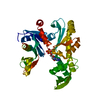
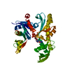
- PDB-8azg: Crystal structure of MreB from Geobacillus stearothermophilus ATC... -
+ Open data

- Basic information
| Entry | Database: PDB / ID: 8azg | ||||||
|---|---|---|---|---|---|---|---|
| Title | Crystal structure of MreB from Geobacillus stearothermophilus ATCC7953 in complex with ATP | ||||||
 Components Components | Cell shape-determining protein MreB | ||||||
 Keywords Keywords | STRUCTURAL PROTEIN / bacterial actin / bacterial cytoskeleton | ||||||
| Function / homology |  Function and homology information Function and homology informationcell morphogenesis / regulation of cell shape / GTP binding / ATP binding / cytoplasm Similarity search - Function | ||||||
| Biological species |   Geobacillus stearothermophilus ATCC 7953 (bacteria) Geobacillus stearothermophilus ATCC 7953 (bacteria) | ||||||
| Method |  X-RAY DIFFRACTION / SYNCHROTRON / MOLECULAR REPLACEMENT / Resolution: 2.29 Å X-RAY DIFFRACTION / SYNCHROTRON / MOLECULAR REPLACEMENT / Resolution: 2.29 Å | ||||||
 Authors Authors | Li de la Sierra-Gallay, I. / Mao, W. | ||||||
| Funding support |  France, 1items France, 1items
| ||||||
 Citation Citation | Journal: Elife / Year: 2023 Title: On the role of nucleotides and lipids in the polymerization of the actin homolog MreB from a Gram-positive bacterium. Authors: Mao, W. / Renner, L.D. / Cornilleau, C. / Li de la Sierra-Gallay, I. / Afensiss, S. / Benlamara, S. / Ah-Seng, Y. / Van Tilbeurgh, H. / Nessler, S. / Bertin, A. / Chastanet, A. / Carballido-Lopez, R. | ||||||
| History |
|
- Structure visualization
| Structure viewer | Molecule: MolmilJmol/JSmol |
|---|
- Downloads & links
-Download
| PDBx/mmCIF format | 8azg.cif.gz | 96.5 KB | Display | PDBx/mmCIF format |
|---|---|---|---|---|
| PDB format | pdb8azg.ent.gz | 56.8 KB | Display | PDB format |
| PDBx/mmJSON format | 8azg.json.gz | Tree view | PDBx/mmJSON format | |
| Others |  Other downloads Other downloads |
-Validation report
| Arichive directory | https://data.pdbj.org/pub/pdb/validation_reports/az/8azgftp://data.pdbj.org/pub/pdb/validation_reports/az/8azg | HTTPS FTP |
|---|
-Related structure data
| Related structure data | 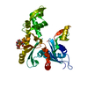 7zptSC 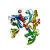 7zpuC S: Starting model for refinement C: citing same article ( |
|---|---|
| Similar structure data |
-Links
 PDBj
PDBj- Assembly
| Deposited unit | 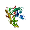
| ||||||||||||
|---|---|---|---|---|---|---|---|---|---|---|---|---|---|
| 1 |
| ||||||||||||
| Unit cell |
|
-Components
| #1: Protein | Mass: 37405.078 Da / Num. of mol.: 1 Source method: isolated from a genetically manipulated source Source: (gene. exp.) Geobacillus stearothermophilus ATCC 7953 (bacteria)Gene: mreB, B4109_2230, B4114_2117, GS458_2390 / Production host: | ||||
|---|---|---|---|---|---|
| #2: Chemical | ChemComp-ATP /   Mass: 507.181 Da / Num. of mol.: 1 / Source method: obtained synthetically / Formula: C10H16N5O13P3 / Feature type: SUBJECT OF INVESTIGATION / Comment: ATP, energy-carrying molecule*YM Mass: 507.181 Da / Num. of mol.: 1 / Source method: obtained synthetically / Formula: C10H16N5O13P3 / Feature type: SUBJECT OF INVESTIGATION / Comment: ATP, energy-carrying molecule*YM | ||||
| #3: Chemical |   Mass: 92.094 Da / Num. of mol.: 3 / Source method: obtained synthetically / Formula: C3H8O3 Mass: 92.094 Da / Num. of mol.: 3 / Source method: obtained synthetically / Formula: C3H8O3#4: Water | ChemComp-HOH / |  Mass: 18.015 Da / Num. of mol.: 81 / Source method: isolated from a natural source / Formula: H2O Mass: 18.015 Da / Num. of mol.: 81 / Source method: isolated from a natural source / Formula: H2OHas ligand of interest | Y | |
-Experimental details
-Experiment
| Experiment | Method: X-RAY DIFFRACTION / Number of used crystals: 1 |
|---|
- Sample preparation
| Crystal | Density Matthews: 2.94 Å3/Da / Density % sol: 58.15 % |
|---|---|
| Crystal grow | Temperature: 293 K / Method: vapor diffusion, sitting drop Details: PEG 8000, glycerol, potassium phosphate, magnesium chloride |
-Data collection
| Diffraction | Mean temperature: 95 K / Serial crystal experiment: N |
|---|---|
| Diffraction source | Source: SYNCHROTRON / Site: SOLEIL / Beamline: PROXIMA 1 / Wavelength: 0.978565 Å |
| Detector | Type: DECTRIS EIGER X 16M / Detector: PIXEL / Date: Nov 9, 2019 |
| Radiation | Protocol: SINGLE WAVELENGTH / Monochromatic (M) / Laue (L): M / Scattering type: x-ray |
| Radiation wavelength | Wavelength: 0.978565 Å / Relative weight: 1 |
| Reflection | Resolution: 2.29→43.88 Å / Num. obs: 19996 / % possible obs: 99.7 % / Redundancy: 13.13 % / Biso Wilson estimate: 48.1 Å2 / CC1/2: 1 / Rrim(I) all: 0.146 / Net I/σ(I): 12.41 |
| Reflection shell | Resolution: 2.29→2.43 Å / Num. unique obs: 3130 / CC1/2: 0.84 / Rrim(I) all: 1.41 |
- Processing
| Software |
| ||||||||||||||||||||||||||||||||||||||||||||||||||||||||
|---|---|---|---|---|---|---|---|---|---|---|---|---|---|---|---|---|---|---|---|---|---|---|---|---|---|---|---|---|---|---|---|---|---|---|---|---|---|---|---|---|---|---|---|---|---|---|---|---|---|---|---|---|---|---|---|---|---|
| Refinement | Method to determine structure: MOLECULAR REPLACEMENT Starting model: 7ZPT Resolution: 2.29→42.47 Å / SU ML: 0.2779 / Cross valid method: FREE R-VALUE / σ(F): 1.35 / Phase error: 28.9527 Stereochemistry target values: GeoStd + Monomer Library + CDL v1.2
| ||||||||||||||||||||||||||||||||||||||||||||||||||||||||
| Solvent computation | Shrinkage radii: 0.9 Å / VDW probe radii: 1.1 Å / Solvent model: FLAT BULK SOLVENT MODEL | ||||||||||||||||||||||||||||||||||||||||||||||||||||||||
| Displacement parameters | Biso mean: 58.48 Å2 | ||||||||||||||||||||||||||||||||||||||||||||||||||||||||
| Refinement step | Cycle: LAST / Resolution: 2.29→42.47 Å
| ||||||||||||||||||||||||||||||||||||||||||||||||||||||||
| Refine LS restraints |
| ||||||||||||||||||||||||||||||||||||||||||||||||||||||||
| LS refinement shell |
|
