 Movie
Movie Controller


+ Open data

- Basic information
| Entry | Database: PDB / ID: 8ayt | ||||||
|---|---|---|---|---|---|---|---|
| Title | Crystal structure of SUDV VP40 W95A mutant | ||||||
 Components Components | Matrix protein VP40 | ||||||
 Keywords Keywords | VIRAL PROTEIN / Ebola virus / SUDV / VP40 / matrix protein / dimer | ||||||
| Function / homology |  Function and homology information Function and homology informationhost cell endomembrane system / host cell late endosome membrane / viral budding via host ESCRT complex / structural constituent of virion / symbiont-mediated suppression of host innate immune response / ribonucleoprotein complex / host cell plasma membrane / virion membrane / RNA binding / membrane Similarity search - Function | ||||||
| Biological species |  Sudan ebolavirus Sudan ebolavirus | ||||||
| Method |  X-RAY DIFFRACTION / SYNCHROTRON / MOLECULAR REPLACEMENT / molecular replacement / Resolution: 1.9 Å X-RAY DIFFRACTION / SYNCHROTRON / MOLECULAR REPLACEMENT / molecular replacement / Resolution: 1.9 Å | ||||||
 Authors Authors | Werner, A.-D. / Steinchen, W. / Werel, L. / Kowalski, K. / Essen, L.-O. / Becker, S. | ||||||
| Funding support |  Germany, 1items Germany, 1items
| ||||||
 Citation Citation | Journal: To Be Published Title: Crystal structure of SUDV VP40 W95A mutant Authors: Werner, A.-D. / Becker, S. | ||||||
| History |
|
- Structure visualization
| Structure viewer | Molecule: MolmilJmol/JSmol |
|---|
- Downloads & links
-Download
| PDBx/mmCIF format | 8ayt.cif.gz | 131.9 KB | Display | PDBx/mmCIF format |
|---|---|---|---|---|
| PDB format | pdb8ayt.ent.gz | 84.8 KB | Display | PDB format |
| PDBx/mmJSON format | 8ayt.json.gz | Tree view | PDBx/mmJSON format | |
| Others |  Other downloads Other downloads |
-Validation report
| Summary document | 8ayt_validation.pdf.gz | 415.7 KB | Display | wwPDB validaton report |
|---|---|---|---|---|
| Full document | 8ayt_full_validation.pdf.gz | 416.6 KB | Display | |
| Data in XML | 8ayt_validation.xml.gz | 11.6 KB | Display | |
| Data in CIF | 8ayt_validation.cif.gz | 16 KB | Display | |
| Arichive directory | https://data.pdbj.org/pub/pdb/validation_reports/ay/8aytftp://data.pdbj.org/pub/pdb/validation_reports/ay/8ayt | HTTPS FTP |
-Related structure data
| Related structure data |  4ld8S S: Starting model for refinement |
|---|---|
| Similar structure data |
-Links
 PDBj
PDBj
- Assembly
| Deposited unit | 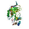
| ||||||||||
|---|---|---|---|---|---|---|---|---|---|---|---|
| 1 | 
| ||||||||||
| Unit cell |
|
-Components
| #1: Protein | Mass: 32488.484 Da / Num. of mol.: 1 Source method: isolated from a genetically manipulated source Source: (gene. exp.) Sudan ebolavirus / Gene: VP40, DF49_53412gpVP40, DH33_45403gpVP40, SEBOVgp3 / Production host:  |
|---|---|
| #2: Water | ChemComp-HOH /  Mass: 18.015 Da / Num. of mol.: 121 / Source method: isolated from a natural source / Formula: H2O Mass: 18.015 Da / Num. of mol.: 121 / Source method: isolated from a natural source / Formula: H2O |
-Experimental details
-Experiment
| Experiment | Method: X-RAY DIFFRACTION / Number of used crystals: 1 |
|---|
- Sample preparation
| Crystal | Density Matthews: 2.11 Å3/Da / Density % sol: 41.81 % |
|---|---|
| Crystal grow | Temperature: 293 K / Method: vapor diffusion, sitting drop Details: 0.5 M potassium chloride 50 mM MOPS 12% (w/v) PEG4000 20% (w/v) glycerol |
-Data collection
| Diffraction | Mean temperature: 100 K / Serial crystal experiment: N |
|---|---|
| Diffraction source | Source: SYNCHROTRON / Site: SLS  / Beamline: X06SA / Wavelength: 1.000009 Å / Beamline: X06SA / Wavelength: 1.000009 Å |
| Detector | Type: DECTRIS EIGER X 16M / Detector: PIXEL / Date: Oct 26, 2019 |
| Radiation | Protocol: SINGLE WAVELENGTH / Monochromatic (M) / Laue (L): M / Scattering type: x-ray |
| Radiation wavelength | Wavelength: 1.000009 Å / Relative weight: 1 |
| Reflection | Resolution: 1.9→48.6 Å / Num. obs: 22067 / % possible obs: 99.1 % / Redundancy: 3.8 % / Biso Wilson estimate: 33.51 Å2 / CC1/2: 0.99 / Net I/σ(I): 10.7 |
| Reflection shell | Resolution: 1.9→1.94 Å / Redundancy: 3.6 % / Mean I/σ(I) obs: 2.5 / Num. unique obs: 1406 / CC1/2: 0.912 / % possible all: 98.2 |
-Phasing
| Phasing | Method: molecular replacement |
|---|
- Processing
| Software |
| ||||||||||||||||||||||||||||||||||||||||||||||||||||||||||||||||||||||||||||||||||||||||||||||||||||
|---|---|---|---|---|---|---|---|---|---|---|---|---|---|---|---|---|---|---|---|---|---|---|---|---|---|---|---|---|---|---|---|---|---|---|---|---|---|---|---|---|---|---|---|---|---|---|---|---|---|---|---|---|---|---|---|---|---|---|---|---|---|---|---|---|---|---|---|---|---|---|---|---|---|---|---|---|---|---|---|---|---|---|---|---|---|---|---|---|---|---|---|---|---|---|---|---|---|---|---|---|---|
| Refinement | Method to determine structure: MOLECULAR REPLACEMENT Starting model: 4LD8 Resolution: 1.9→45.64 Å / SU ML: 0.1813 / Cross valid method: FREE R-VALUE / σ(F): 1.38 / Phase error: 23.1826 Stereochemistry target values: GeoStd + Monomer Library + CDL v1.2
| ||||||||||||||||||||||||||||||||||||||||||||||||||||||||||||||||||||||||||||||||||||||||||||||||||||
| Solvent computation | Shrinkage radii: 0.9 Å / VDW probe radii: 1.11 Å / Solvent model: FLAT BULK SOLVENT MODEL | ||||||||||||||||||||||||||||||||||||||||||||||||||||||||||||||||||||||||||||||||||||||||||||||||||||
| Displacement parameters | Biso mean: 49.41 Å2 | ||||||||||||||||||||||||||||||||||||||||||||||||||||||||||||||||||||||||||||||||||||||||||||||||||||
| Refinement step | Cycle: LAST / Resolution: 1.9→45.64 Å
| ||||||||||||||||||||||||||||||||||||||||||||||||||||||||||||||||||||||||||||||||||||||||||||||||||||
| Refine LS restraints |
| ||||||||||||||||||||||||||||||||||||||||||||||||||||||||||||||||||||||||||||||||||||||||||||||||||||
| LS refinement shell |
| ||||||||||||||||||||||||||||||||||||||||||||||||||||||||||||||||||||||||||||||||||||||||||||||||||||
| Refinement TLS params. | Method: refined / Refine-ID: X-RAY DIFFRACTION
| ||||||||||||||||||||||||||||||||||||||||||||||||||||||||||||||||||||||||||||||||||||||||||||||||||||
| Refinement TLS group |
|
